Physiological Monitoring Subsystem
Visualization and Movement Detection
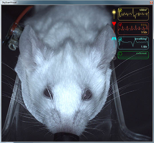
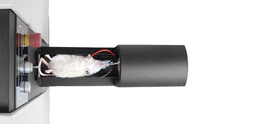
The physiological monitoring sub-system includes video monitoring of an animal with real-time movement detection, ECG and breathing detection, and temperature stabilization. A colour TV-camera (5 Mp in SKYSCAN 1278) is mounted above the animal bed and equipped with white LED illumination to produce a real-time image of the animal during the scan. The software analyses the video stream from a user selected area of the image, which the operator can position on a part of the animal body where breathing movement is visible. These movements are converted into a movement waveform to provide breathing timemarks for time-resolved micro-CT reconstruction.
The face mask on the animal bed is connected to an air/gas flow sensor for direct breathing detection. The ECG electrodes in the animal cassette are connected to a sensitive ECG amplifier. Both breathing and ECG signals are digitized, sent to computer and displayed as real-time profiles on-screen. An operator can select individual gain and threshold for each signal to optimize generation of timemarks.
The monitoring also includes temperature stabilization by heated airflow, which maintains the scanned animal at a selected temperature, to prevent cooling of the animal under anaesthesia.
4D Time-Resolved Microtomography
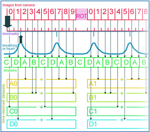
Physiological monitoring creates reference time-marks for time-resolved reconstruction of heart and lung dynamics, invented by Bruker microCT. In 4D (time-resolve) scanning mode, multiple projection images taken at each gantry angular position are sorted post-scan into breathing or heart time bins using recorded physiological monitoring timemarks. Such sorting creates pseudo-static sets of projections, which are reconstructed as separate datasets and produce 3D sets of results corresponding to different phases of the cardiac or respiratory cycle.
Our visualization program loads all reconstructed datasets and allows scrolling in XYZ dimension across the reconstructed volume as well as in the time-dimension to demonstrate dynamics of heart or lung movements in sharp reconstructed images minimally affected by movement artifacts. Because all acquired data are sorted after the acquisition process, respiratory and cardiac cycles can be visualized by sorting according to timemarks from physiological monitoring without rescanning the animal.
All-Carbon ECG Electrodes
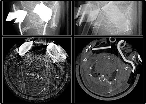
The animal cassettes include special cables and clip electrodes to detect ECG signals by a sensitive amplifier integrated in the physiological monitoring sub-system. The ECG connections use ECG wiring and electrodes specially developed by Bruker microCT which contain no metal parts. The wires and electrodes employ advanced carbon-fiber conductive parts with X-ray absorption similar to that of animal tissues for uncompromised image quality.