Application Note: Deep Brain Imaging with nVista™
Discover the benefits of deep brain imaging with nVista™
In this application highlight, readers can expect to learn more about the nVista system, including how it works and examples of published data. Explore how to record and process calcium imaging data to uncover neural circuitry and behavioral dynamics in freely behaving animals.
Readers can expect to learn more about:
- Imaging neuronal activity at single-cell resolution in deep brain regions such as the hypothalamus, basal forebrain, and laterodorsal tegmental nucleus with nVista™
- How calcium imaging during naturalistic behaviors provides insights into functions like arousal, reward, and cognition
- Advanced data acquisition and analysis tools for Inscopix miniscopes
- Experimental workflow for viral injection, lens probe implantation, and nVista installation

Introduction
Deep brain regions like the hypothalamus (LH), basal forebrain (BF) and laterodorsal tegmental nucleus (LDT) regulate multiple essential physiological functions such as emotion, reward, energy homeostasis and arousal state; and impairments to these areas often result in cognitive deficits1-3. These nuclei are deep within the brain (Figure 1) and contain a diversity of cell types. Imaging tools to access these structures in a cell type-specific manner will greatly empower studies designed to elucidate their neural circuitry and behavioral function(s).

In vivo Ca2+ imaging during freely moving behavior
In vivo Ca2+ imaging allows scientists to capture neural activity at single cell resolution within specified cell types. nVista microscopy is a practical way to conduct Ca2+ imaging during active behavior, and capture Ca2+ dynamics repeatedly over time from the same neural populations during various behaviors. Several in vivo Ca2+ imaging studies5-8 of deep brain regions (e.g. LH, BF and LDT) have been published using the Inscopix nVista system. nVista imaging provides new ways to study spatial coding, ensemble dynamics during active behavior as well as assess drug effects on brain function and behavior.
nVista materials and supplies
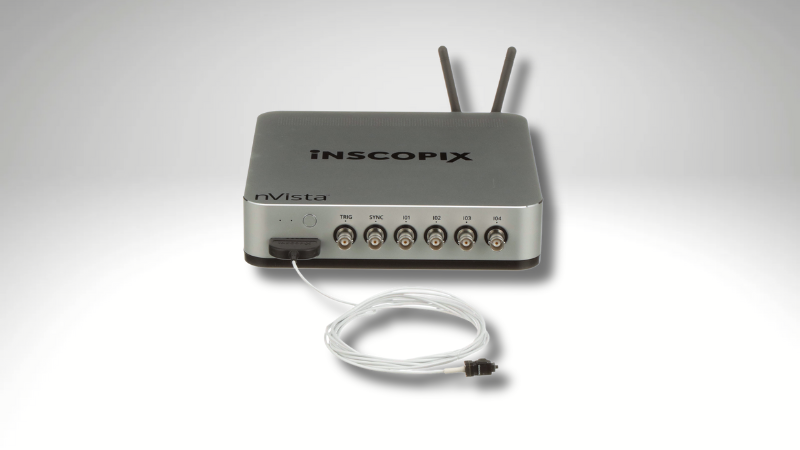
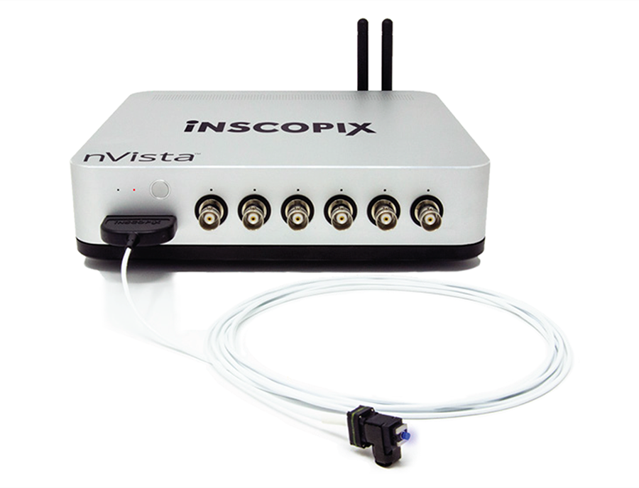
The nVista imaging system (Figure 2) is composed of miniaturized fluorescence microscope, data acquisition box, Inscopix data acquisition and data processing softwares, and hardware accessories. In vivo imaging in deep brain regions in mouse can now be achieved when the nVista system is paired with our implantable lens probes4-8.
Other materials needed:
- Stereotax with manipulator
- Microinjection pump
- Approved rodent survival surgery tools and anesthetics
- Dental cement or cyanoacrylate adhesive
- GCaMP virus
- Wildtype mice
In this application highlight, we will outline a brief synopsis of published methods and results showcasing nVista calcium imaging of the BF of freely behaving mice6.
General experimental workflow*
A. Viral injection: target deep brain region of interest with Ca2+ indicator
- Obtain the optimal virus, brain coordinates, volume and injection rate to label your cell type of interest. Confirm viral expression and location with histology.
- Prepare animal for standard survival surgery procedures.
- Inject virus into target brain region with the aid of a stereotax and micropump.
- Suture skin and conduct postoperative recovery procedures.
- Wait approximately 1-2 weeks for viral expression.
B. Enable optical access: implant the lens probe
- Sterilize the lens probe and prepare animal for standard survival stereotaxic surgery.
- Open a craniotomy large enough to permit lens probe access.
- Use the stereotax manipulator arm to securely hold the lens probe, with the imaging side facing the target brain region.
- Slowly lower the lens probe through the craniotomy into the brain until the proper depth is reached. Aspiration may be required.
- Use cyanoacrylate adhesive to fix the lens probe securely to the skull. Use dental cement to create a cranial cap to protect the lens probe and implant site. Ensure that the imaging face of the lens probe is clean and protected.
- Return the animal to a clean home cage. Administer postoperative analgesics (per your institution’s guidelines) and allow for 1-2 weeks of recovery.
C. Install nVista: securing the baseplate docking system
- Prepare animal for standard stereotaxic surgery (note: this step is not an invasive procedure).
- Use stereotax and gripper arm to hold the nVista microscope with attached baseplate above implanted lens probe imaging face. Ensure the objective lens and the lens probe imaging face are parallel.
- Connect the nVista system to a computer, and start the Inscopix Data Acquisition Software. Turn on the nVista microscope LED.
- Observe brain tissue using Inscopix Data Acquisition Software while advancing the nVista objective towards the imaging face of the lens via the stereotax. When tissue is in focus through the lens probe, the nVista microscope is at the proper location for optimal focus.
- Use adhesive to cement the baseplate at this optimal location. Carefully adhere the baseplate only (not the microscope).
- Once the adhesive and baseplate are securely fixed to the skull, remove the nVista microscope, and put on a baseplate cover.
- Return the mouse to its homecage for recovery from anesthesia.
D. Acquire in vivo Ca2+ imaging data
- Plug in the nVista system to the computer. Ensure there is plenty of memory and data storage space.
- Briefly anesthetize or awake restrain the animal, remove the baseplate cover, and attach the nVista microscope.
- Place animal with attached nVista microscope in the behavioral arena of choice. Allow the animal to recover briefly from anesthesia or handling, and begin experiment!
* Abbreviated from detailed experimental and surgical methods in accordance with institution’s guidelines.

Select deep brain publications

LEARN MORE:
Data analysis and results
Imaging neuronal activity during spontaneous behaviors8
Harrison et al. (2016) examined the activity patterns of BF neurons during spontaneous behaviors in mice fitted with the integrated fluorescence microscope (nVista). Spontaneous behaviors (e.g. sitting, eating/grooming, moving or running) were recorded for 25-50 min using a webcam and scored manually (Figure 4).


Processing Ca2+ imaging data
After image acquisition, Harrison and colleagues8 processed the imaging data using Inscopix Data Processing Software. This software is designed to preprocess the raw imaging data and extract Ca2+ dynamics of individual cells and allows researchers to correct for brain motion, calculate ΔF/F, and denoise images, among other manipulations. In addition, this software performs PCA/ICA analysis that allows for the detection of cellular events followed by the extraction of independent Ca2+ signals and sorts them into a relevant context (Figure 5).
Discussion
The ability to study the dynamics of neural circuitry and behavioral states in freely behaving animals is vital to gaining a better understanding of the function of deep brain regions in health and disease.
High-throughput in vivo calcium imaging at single cell resolution with the nVista system paired with the lens probes opens new possibilities in the mouse brain and allows researchers to ask new questions about how these various deep brain regions modulate essential functions such as arousal state, feeding behavior, reward and cognition, in order to address how these neural circuits become perturbed in CNS disease.
References
- Brown et al. Control of sleep and wakefulness. Physiol Rev (2012)
- Ballinger et al. Basal forebrain cholinergic circuits and signaling in cognition and cognitive decline. Neuron (2016)
- Stuber and Wise. Lateral hypothalamic circuits for feeding and reward. Nat Neurosci (2016)
- Resendez et al. Visualization of cortical, subcortical and deep brain neural circuit dynamics during naturalistic mammalian behavior with headmounted microscopes and chronically implanted lenses. Nat Prot (2016)
- Betley et al. Neurons for hunger and thirst transmit a negative-valence teaching signal. Nature (2015)
- Jennings et al. Visualizing hypothalamic network dynamics for appetitive and consummatory behaviors. Cell (2015)
- Cox et al. Calcium imaging of sleep-wake related neuronal activity in the dorsal pons. Nat Comm (2016)
- Harrison et al. Calcium imaging of basal forebrain activity during innate and learned behaviors. Front Neural Circuits (2016)
- Douglass et al. Central amygdala circuits modulate food consumption through a positive-valence mechanism. Nat Neurosci (2017)
- Li et al. Neuronal representation of social information in the medial amygdala of awake behaving mice. Cell (2017)
- Remedios et al. Social behaviour shapes hypothalamic neural ensemble representations of conspecific sex. Nature (2017)
- Ryan et al. Oxytocin-receptor-expressing neurons in the parabrachial nucleus regulate fluid intake. Nat Neurosci (2017)
- Chen et al. A hypothalamic switch for REM and Non-REM sleep. Neuron (2018)
- da Silva et al. Dopamine neuron activity before ction initiation gates and invigorates future movements. Nature (2018)
- Campos et al. Encoding of danger by parabrachial CGRP neurons. Nature (2018)
- Markowitz et al. The striatum organizes 3D behavior via moment-to-moment action selection. Cell (2018)