
AFM – Delivering Profound Insights in Microbiology
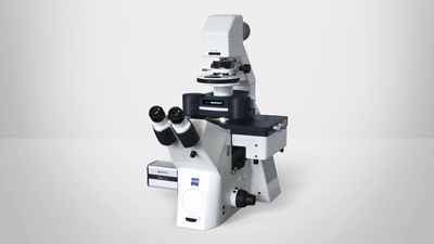
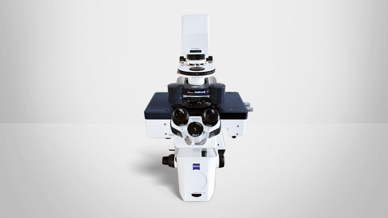
Unravel the 3D Architecture of Cell Membranes with BioAFM
During this webinar, Dr. Laia Pasquina Lemonche presents her work using atomic force microscopy to image cell walls of bacteria at a high resolution. The webinar will also include a live demonstration from Berlin, showcasing the capabilities of Bruker’s NanoWizard BioAFM platform.
Presenter’s Abstract
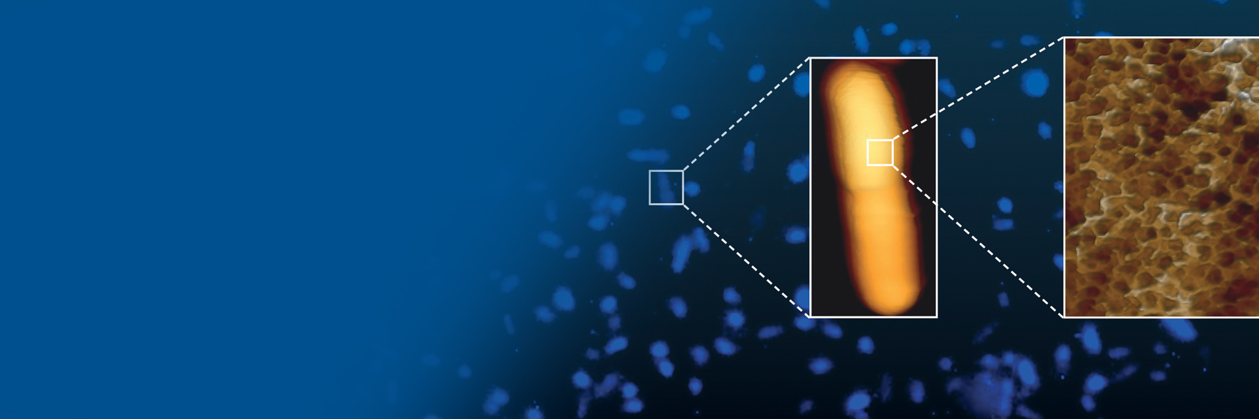
The cell wall of bacteria is mainly composed of a disordered polymer called peptidoglycan. As this is a non-crystalline material, its three-dimensional architecture cannot be determined with the traditional techniques often used in microbiology, such as cryo-EM or NMR. For this reason, the Hobbs and Pasquina groups at the University of Sheffield have, for many years, been using AFM to decipher the three-dimensional architecture of this un-ordered material. Using Bruker’s atomic force microscopes, they have been able to push the imaging resolution of this highly deformable biological material to unprecedented levels. In my talk, I will share AFM methods and present landmark studies.
Find out more about the technology featured in this webinar or our other solutions for BioAFM:
Guest Speaker

Dr. Laia Pasquina Lemonche, The University of Sheffield, UK
Dr. Laia Pasquina Lemonche is an Early-Career-Award Wellcome Trust Fellow. She has a degree in Physics and received a Master’s in Nanobiotechnology from the Autonomous University of Barcelona (UAB) in 2016. In 2020, she obtained a PhD in Biophysics from the University of Sheffield where she focused on deciphering the molecular architecture of gram-positive bacterial cell walls using AFM. She went on to become a Research Associate in Professor Jamie Hobbs’ group until 2024, pushing the limits of resolution of biological material. Currently, her group focuses on using correlative AFM and STORM to study Streptococcus pneumoniae peptidoglycan and developing novel software approaches for microscopy image analysis.