
NMR Reveals New Dimension to p53’s Cancer Role
p53 is well-known for its cancer-preventive role in healthy cells and, when faulty, its near-universal role in the growth of cancer cells. But, despite being uncovered several decades ago, we are still making discoveries about the way this tumor suppressor gene works, which could lead to new drug types to treat the disease.
Under usual circumstances, p53 prevents cells growing uncontrollably by activating a process known as apoptosis, or programmed cell death. This means that when cells are no longer needed or their DNA becomes damaged, they can effectively self-destruct. In cancer, this process – known as the p53 pathway – can become inactivated, allowing cancerous cells to continue to multiply.
Scientists are familiar with p53 as a transcription factor – a protein that works within the nucleus to activate other genes. These genes then produce proteins which ultimately activate the signaling cascade within a cell that leads to its death.
But, around ten years ago, research began to show that p53 is more than just a transcription factor. In fact, it is able to activate apoptosis independently in the cytosol, even in cells that have no nucleus. In 2004, Chipuk et al published research showing how it did so: p53 in the cytosol can directly activate BAX – a key player in the p53 pathway.
However, it was not until recently, with the assistance of nuclear magnetic resonance (NMR) spectroscopy, that the researchers were able to show the exact mechanism by which the protein can do this, and the findings could lead to new drug discoveries for cancer.
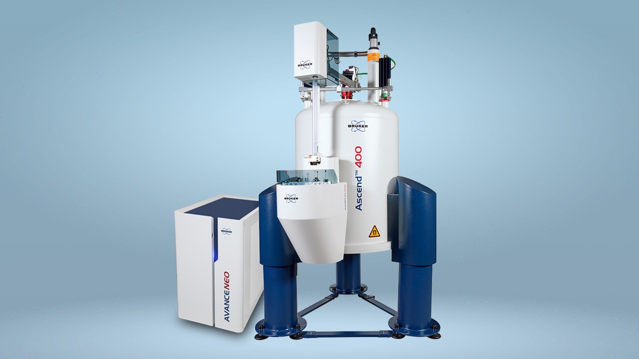
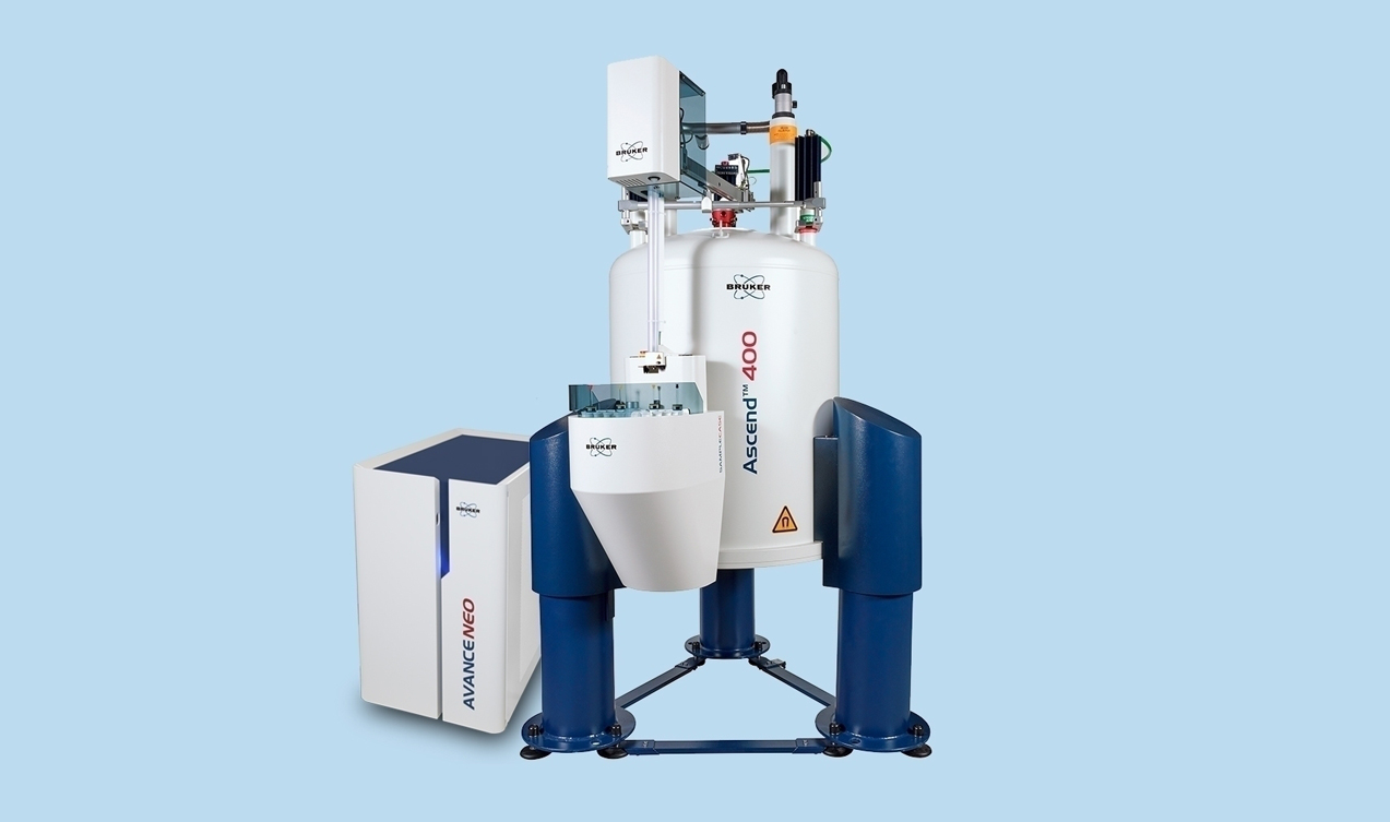
Using a Bruker Avance spectrometer with cryoprobes and Bruker TopSpin software, the researchers looked for chemical shift perturbations (CSPs) when BAX was added to radioactively labeled fragments of p53. This revealed “satellite” peaks for the resonance of a particular proline amino acid in p53. The researchers established that this amino acid was undergoing cis-trans isomerization – switching from its usual trans state into its less biologically common cis state.
Then, by adding fragments of p53 to radioactively labeled BAX they were able to show that two distinct regions of p53 were binding independently to BAX. This was a surprise, the researchers say, as it is different to the established way in which BAX is known to be activated.
By titrating p53 fragments to radioactively labeled BAX, the team were able to observe the kinetics of this binding which drew the findings together: the conformational change of proline was essential for the activation of BAX by p53.
The researchers also used this NMR data to create a molecular model, revealing the precise mechanism in 3D form.
As well as furthering our understanding of apoptosis and fundamental cancer biology, the researchers say that the finding could lead to new drug discoveries through the development of molecules that trigger this newly discovered BAX-dependent apoptosis.
References
- Bradley D. (2015) NMR reveals cancer clue: p53 activity. Available at: http://www.spectroscopynow.com/nmr/details/ezine/14f25d494fb/NMR-reveals-cancer-clue-p53-activity.html. Date accessed: 06 September 2015.
- Chipuk JE, et al. Direct activation of Bax by p53 mediates mitochondrial membrane permeabilization and apoptosis. Science 2004; 303: 1010-1014.
- Chipuk JE, et al. Pharmacological activation of p53 elicits Bax-dependent apoptosis in the absence of transcription. Cancer Cell 2003; 4: 371-381.
- Follis AV, et al. The DNA-binding domain mediates both nuclear and cytosolic functions of p53. Nature Structural & Molecular Biology 2014; 21: 535-543.
- Follis AV, et al. Pin1-Induced Proline Isomerization in Cytosolic p53 Mediates BAX Activation and Apoptosis. Molecular Cell 2015; 59: 677-684.
- Vogelstein B (2010). p53: The most frequently altered gene in human cancers. Available at: https://www.nature.com/scitable/topicpage/p53-the-most-frequently-altered-gene-in-14192717. Date accessed: 6th September 2015.


