Putting the Cell-Cycle Clock into Organoid Phenotyping: From CALIPERS Cardiac Organoids to Lumenoids and Gastruloids
See how cell-cycle dynamics shape organoid development
In this presentation, Francesco S. Pasqualini (University of Pavia) presents CALIPERS, a live-cell imaging framework developed to study how cell-cycle dynamics influence tissue development, organization, and function. Through applications in cardiac organoids, lumenoids, and gastruloids, he demonstrates how cell proliferation and cell cycle state can be connected to developmental and functional outcomes.
Topics covered include:
- Tracking cell-cycle dynamics during organoid development and maturation
- Monitoring tissue organization and functional development in cardiac organoids
- Applying live imaging approaches to lumenoids, gastruloids, and regenerative biology studies
Putting the Cell-Cycle Clock into Organoid Phenotyping: From CALIPERS Cardiac Organoids to Lumenoids and Gastruloids
Cell-cycle state is a hidden coordinate in live-cell phenotyping, especially in stem-cell-derived 3D tissues where proliferation, lineage commitment, morphogenesis, and functional maturation unfold together.
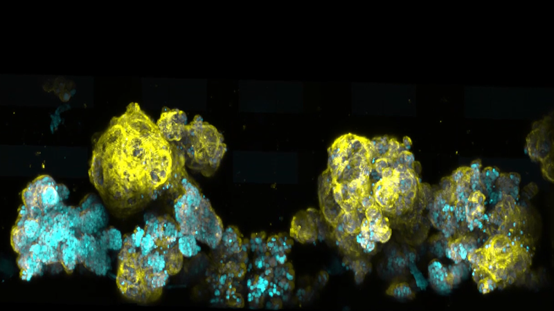
I will present CALIPERS, our recently published Nature Communications framework for cell-cycle-aware live-cell imaging in phenotyping experiments and regeneration studies. CALIPERS combines a spectrally redesigned FUCCI reporter, continuous cell-cycle inference, and compatible structural and functional biosensors in human cell and hiPSC models. In cardiac organoids, it tracks cell-cycle exit, tissue compaction, calcium onset, and distinguishes productive cardiomyocyte proliferation from multinucleation and endoreplication.
I will then discuss how CALIPERS hiPSC reporter cells are being extended into Lumenoids and Gastruloids to link cell-cycle dynamics to tissue organization and to enable live light-sheet phenotyping in developmental and regenerative contexts.
Featured Technologies:
Contact:
Find out more about the technology featured in this webinar or our other solutions for light-sheet microscopy:
Speaker

Francesco S. Pasqualini
Associate Professor of Industrial Bioengineering, Principal Investigator @ Synthetic Physiology Laboratory, Department of Civil Engineering and Architecture, University of Pavia