Development and Embryology
Developmental biology and embryology benefit from the ability to acquire different data types with light-sheet microscopy. For example, 3D data stacks can be acquired at single time points, and when imaging multiple samples, one can create developmental templates and understand developmental plasticity.
Using time-lapse imaging, structural and functional studies, and calcium imaging and single-cell tracking allows insights into dynamic processes occurring during development. Fixed and cleared tissue samples enable researchers to simultaneously explore development at both the cellular and organism levels.
By employing a variety of models and experimental approaches, scientists can gain a comprehensive understanding of intricate biological processes at play and complementary insights into developmental biology.
Light-sheet microscopy stands out as an imaging technique with:
- A gentle approach that allows imaging of fragile samples
- Low phototoxicity enabling long time-lapse imaging with healthy samples
- Fast imaging speed for imaging multiple samples in very short timeframes, ensuring sample comparability
Applications include:
- Cell tracking and morphogenesis
- Cell differentiation, maturation, and specialization
- Developmental genetics, signalling pathways, and drug screening
Case Study – Cell Tracking
Cell tracking and the computational analysis of this cellular behavior provide insights into the dynamic processes governing tissue formation and organogenesis. Researchers can decipher the mechanisms and processes of cell migration, proliferation, differentiation, and signaling by monitoring individual cells over time. This enables the construction of spatiotemporal maps of embryonic development, cellular responses, and tissue regeneration.
Ultimately, cell tracking and the computational analysis of these data can contribute to our understanding of how organisms form and develop.
Cell Tracking in Drosophila Embryo Development
Cell tracking created with arivis Vision4D 3D visualization and analysis software. Imaged on the MuVi SPIM.
Courtesy of:
Celia Smits and Stanislav Y. Shvartsman
Department of Molecular Biology
Princeton University, NJ
USA
Zebrafish Development
Zebrafish imaged on the MuVi SPIM. Stitched from 5 stacks in Imaris, each 340 slices, 16 hour/10min. Fish growth can be observed.
Courtesy of:
Prof. Jingxia Liu
Huazhong Agricultural University
China
Application Data
Whole Organism Development
Studying mouse development holds immense importance in developmental biology for several reasons. Mice share close genetic similarity with humans, which makes them an invaluable model organism to understand fundamental embyrogenesis and organogenesis. Also, insights gained from mouse studies often have direct relevance to human biology and disease and can aid in the discovery of potential therapeutic strategies for various conditions.
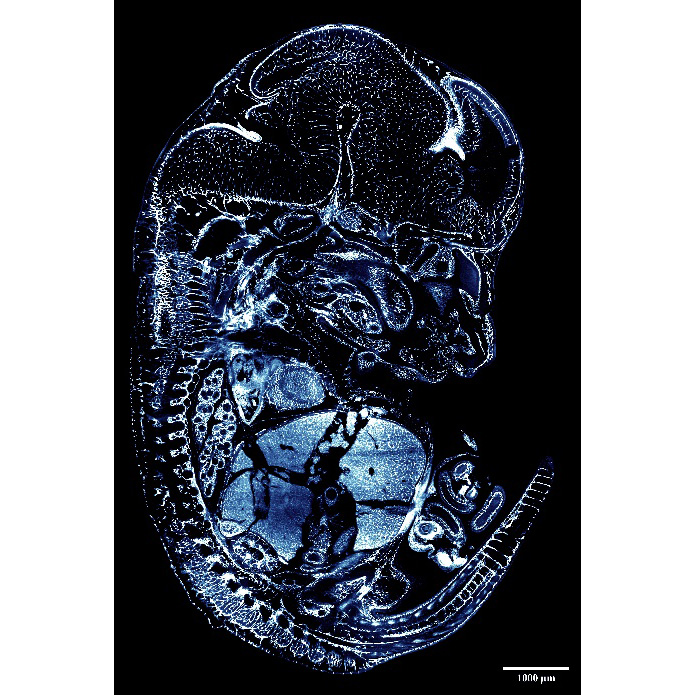
Images acquired with the LCS (Olympus 4x NA 0.28 detection objective) show a single slice of an iDISCO+ cleared E12.5 mouse embryo. Images were rendered in Imaris (Bitplane).
Images courtesy of Sonja Nowotschin, Ying-Yi Kuo, and Kat Hadjantonakis.
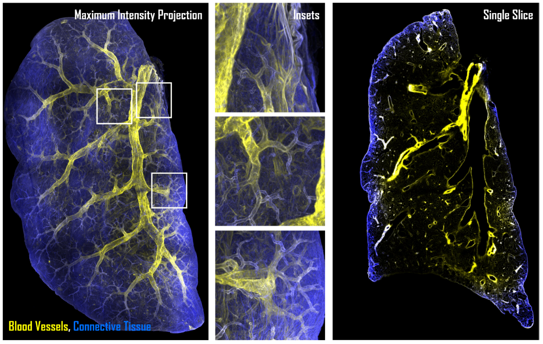
Whole Organ Imaging - Geometric Patterning of Tissues
The combination of tissue clearing and light-sheet imaging can deliver insights into in-toto geometric patterning of tissues, organs, and organisms. This application highlights the intricate branching patterns of mouse lungs and blood vessels.
Zebrafish Vascular Biology
The vascular system transports blood, oxygen, and nutrients through the body, and therefore, plays a pivotal role in embryonic development, health, and disease. Investigating vascular development provides not only insights into biological processes but also the origins of congenital vascular diseases. These insights can potentially pave the way for early diagnosis and intervention.
Moreover, embryonic vascular research has broader implications for regenerative medicine and tissue engineering, as it offers valuable insights into mechanisms of vascular growth and repair.
Vascular Development in Zebrafish
From left to right: the video shows a beating Zebrafish heart imaged at 50 frames/sec, followed by Zebrafish blood vessels (magenta) and red blood cells (yellow) and Zebrafish blood flow imaged at 50 frames/sec. Imaged on the MuVi SPIM.
Courtesy of:
Nadia Mercader & Inés Marques
University of Bern
Bern, Switzerland
Imaging of 3D Cell Cultures
Left: Mouse preimplantation embryos expressing H2B-mCherry. Nuclei tracking from one-cell stage to blastocyst.
Right: Mouse oocytes expressing CENPC-EGFP and H2B-mCherry for kinetochore tracking.
Imaged on the InVi SPIM.
Petr Strnad, et al. (2016). Inverted light-sheet microscope for imaging mouse pre-implantation development. Nature Methods 13, 139-145
Mitosis in HeLa cells stained for histone 2B-mCherry (magenta), GFP-tubulin (green) and GFP-tubulin (white, deconvolved).
Imaged on the InVi SPIM Lattice Pro.
Visualization: Imaris (Bitplane).
Courtesy of:
Sabine Reither
European Molecualr Biology Laboratory (EMBL)
Heidelberg, Germany
Sample: HeLa cells (Neumann et al., Nature. 2010 Apr 1;464(7289):721-7)

Epithelial cell line (BS-C-1) stained for f-actin and chromatin. Image taken with InVi SPIM Lattice Pro.
Courtesy of:
Ulrike Engel
Nikon Imaging Center
University of Heidelberg
Germany
Imaging of Organoids and Spheroids
hESC-derived pancreatic spheres. Imaging on the TruLive3D Imager enables collecting information of several samples in one experiment. Visualization: Imaris (Bitplane)
Courtesy of:
Yung Hae Kim
Graphin-Botton Group, MPI-CBG
Dresden, Germany
Characterization and imaging of stochastic tumorigenesis in mammary organoids. Imaged on the InVi SPIM.
A. Alladin, L. Chaible, L. Garcia del Valle, S. Reither Sabine, M. Loeschinger, M. Wachsmuth, J.K. Hériché, C. Tischer, M, Jechlinger. Tracking cells in epithelial acini by light-sheet microscopy reveals proximity effects in breast cancer initiation. eLife 2020;9:e54066 doi: 10.7554/eLife.54066
Spheroid stained with anti-GFAP (Alexa 488) to label astrocytes and anti-Neurofilament200 (Alexa 555) to label neurons. Imaged on the InVi SPIM.
Courtesy of:
Markus Bruell
AG Leist, University of Konstanz
Konstanz, Germany
Live Imaging of Plants
Transgenic Arabidopsis root expressing nuclear envelope marker imaged with the MuVi SPIM. The comparison of the videos shows the effect of gravity on root growth.
Courtesy of:
Shanjin Huang
Tsinghua University
Beijing, China
Transgenic Arabidopsis root expressing a membrane marker. Imaged with the InVi SPIM.
Courtesy of:
Alexis Maizel
COS, University of Heidelberg
Heidelberg, Germany

Autofluorescence in microalgae. Imaged on the InVi SPIM.
Other Applications
Mammalian tumors (red) growing in zebrafish (green). Imaged on the MuVi SPIM.
Courtesy of:
Dr. Xi Yao and Dr. Zhangzhao Junjie
Model Animal Research Center of Nanjing University
China
Auto-fluorescence in Daphnia imaged on the MuVi SPIM.
Courtesy of:
Ellen Decaestecker and Luc De Meester
KU Leuven
Kortrijk, Belgium
Protein dynamics in C. elegans skeleton. Imaged on the InVi SPIM.
Courtesy of:
Dr. Chai Yongping and Dr. Ou Guangshuo
Tsinghua University
China
Embryogenesis & Developmental Biology
Drosophila Embryo Development
Transgenic line expressing His2Av-mCherry as fluorescent nuclear reporter. The fruit fly embryo was imaged for almost one complete day (4 × 200 slices every 30 seconds). Imaged on the MuVi SPIM.
Courtesy of:
Lars Hufnagel
European Molecular Biology Laboratory (EMBL)
Heidelberg, Germany
Zebrafish Embryonic Development
Zebrafish embryo expressing Histone H2A-GFP imaged every 6 min from late gastrula to 15–17 somite stage. Imaged on the MuVi SPIM.
Courtesy of:
Andres Collazo
Caltech, Pasadena, CA, USA
as well as: Course faculty and participants of the 2017 Zebrafish Course
Marine Biological Laboratory (MBL)
Woods Hole, MA, USA
Drosophila Egg Chambers
Drosophila ovariole stained with phalloidin to label actin (red) found along membranes and the germline ring canals, DAPI (blue) to show the nuclei, and a somatic ring canal marker (green) to label the ring canals in the epithelium.
Courtesy of:
Jasmin Imran Alsous
Schvartsman Lab
Princeton University, Princeton, NJ, USA