Use of Ultra-high Field MRI for In and Ex Vivo Neuroimaging
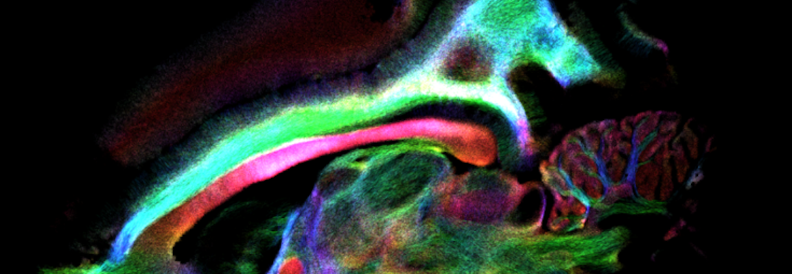
In this webinar, Dr. Mathieu D. Santin from ICM, Paris, France, will describe methods and results obtained for in and ex vivo imaging at ultra-high field in the context of neurodegenerative diseases (demyelinating diseases, Alzheimer disease, multiple sclerosis…). For the investigation of such diseases at UHF Santin primarily implements Multi Gradient Echo (MGE) and 3D-segmented Diffusion EPI techniques. Other innovative studies combining ultrasound and MRI to open the blood brain barrier will be discussed as well. Focused Ultrasound (FUS) are used for instance for cortical activations in primates (Transcranial Focused Ultrasound) but also for non-invasively modulating the visual cortex activity of primates by local ultrasound-induced delivery of an inhibitory neurotransmitter (GABA) : transient disruption of the blood brain barrier (BBB) with FUS coupled with ultrasound contrast agents (UCA).
This webinar took place on October 10th 2019
What to Expect
MR methods for high resolution imaging, diffusion weighted imaging, quantitative susceptibility mapping applied to neurodegenerative diseases; use of Focused-ultrasound combined with MRI studies (cortical activations, blood brain barrier opening…).
Key Topics
- MR methods and results for high resolution imaging
- Applications for neurodegenerative diseases
- Combination of ultrasound and MR techniques
Who Should Attend?
People interested in preclinical MRI, ex vivo MRI, neurodegenerative diseases, highly-resolved anatomical and diffusion MRI, Quantitative Susceptibility Mapping, ultrasound-based MRI studies.
Speakers

Dr. Mathieu D. Santin
Project leader, Neuroimaging platform, CENIR, Brain and Spine Institute (ICM), Paris, France.
After completing his PhD in ultrasound and contrast agents, Mathieu D. Santin performed postdoctorial studies in neuroimaging where he developed new methods based on blood brain barrier opening with ultrasound to monitor amyloid load in mice models. He then joined the Brain and Spine Institute to develop neuroimaging projects.