
Untargeted Biomarker Discovery
MALDI Imaging for untargeted biomarker discovery and classification
Molecular changes originate at the cellular level and solution-based techniques lack sensitivity to capture changes produced from small cell populations.
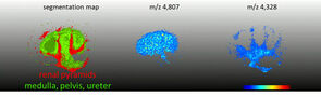
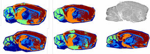
MALDI Imaging of sample cohorts along with SCiLS™ Lab analytical software extracts unique spatial patterns of proteins, lipids or metabolites and correlates them with sample pathology to identify and map putative biomarker ions.
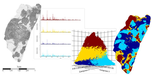
Analyze large sample cohorts for biomarker discovery using molecular profiles and sample metadata to find relevant biomarkers that compliment histology. SCiLS™ Lab is the leading biomarker discovery software for analyzing mass spectrometry imaging with 2D and 3D visualization, as well as sophisticated statistical analysis with just a few mouse clicks, including: automatic spatial segmentation, co-localization analysis, classification model calculation, ROC and component analysis (PCA,pLSA etc).
Bruker’s SCiLS™ Lab analytical software can mine hundreds of thousands of pixels using a variety of statistical tools.
Biomarker discovery protocols can accommodate fresh-frozen or formalin-fixed and paraffin embedded (FFPE) tissues or tissue microarrays (TMAs). Many tissue banks routinely fix biological specimens for long-term storage. Fixation agents act by crosslinking proteins into an insoluble Network, creating an excellent preservation technique. FFPE protocols involve additional processing steps of antigen-retrieval and protein digestion. Numerous publications have demonstrated the utiilty of imaging tryptic peptides from tissue or liberated N-linked glycans.
Hear how Bruker’s MALDI Imaging tools for biomarker discovery are helping Prof. Richard Drake of the Medical University of South Carolina, to gain new insight into the glycosylation pathways of cancer development.
MALDI Guided SpatialOMx®
When your research demands deepest molecular content at the highest spatial context, MALDI Guided SpatialOMx® provides a high definition, label-free overview that merges chemical information with vital morphological context. Click on the link below to learn more about how the revolutionary timsTOF fleX can expand your discovery perspective to encompass proteomics, lipidomics, glycomics, and metabolomics.
Increase lab output with MALDI Imaging
Bruker Daltonics IntelliSlides® now support a brand new workflow which allows for fast, simple, and highly automated set-up of Imaging acquisitions using flexImaging 6.0 and COMPASS® for flex 2.1.
Using the TissueScout to scan barcoded IntelliSlides® allows for fast recognition of the correct sample image in flexImaging as well as alignment of the sample with instrument optics. The new workflow includes checks to confirm instrument performance, and automatic assignment of measurement regions using dedicated algorithms for whole tissue sections and Tissue microarrays (TMAs).
MALDI Imaging is made simple with Bruker Daltonics.
Liteature:
Brochure: Imaging made simple. Streamlined tools for MALDI Imaging experiment set up
App Note: Imaging venom peptides and proteins at high mass resolution, high lateral resolution and high speed using the timsTOF fleX
App Note: The Ease of Use workflow — A simple way to set up MALDI Imaging measurements
Additional information about the efficiency improvements with IntelliSlides® and flexImaging 6.0 can be found in the video:


For Research Use Only. Not for use in clinical diagnostic procedures.