Going Deep: Cross-section Analysis of the Salvator Mundi

The rediscovered Leonardo da Vinci painting, Salvator Mundi, provides a unique window into the materials and techniques used by this master in developing sophisticated visual effects from a relatively limited palette.
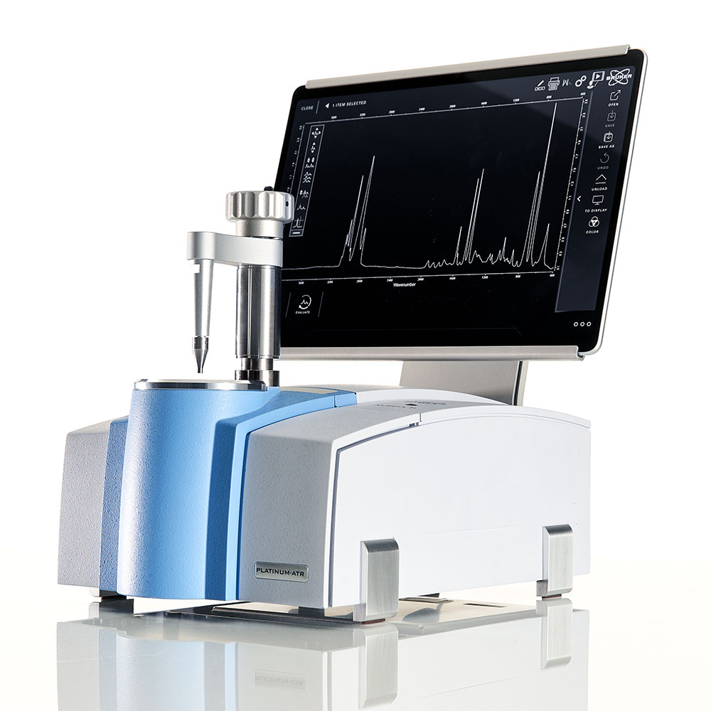
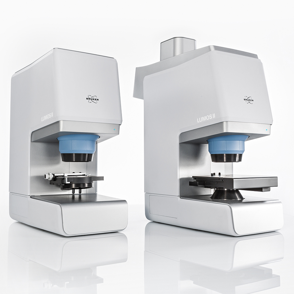
A recent multi-disciplinary study of paint cross-sections obtained from different parts of the painting have allowed unprecedented detail on pigment stratigraphy. Non-destructive analyses were conducted using Fourier Transform Infrared spectroscopy with attenuated total reflection (ATR-FTIR) with Bruker's HYPERION 3000 FTIR Microscope and VERTEX 70 FTIR Spectrometer, and microRaman spectroscopy with Bruker's SENTERRA Raman microscope, along with a number of other complementary techniques. Working in detail with cross-sections carefully removed from intact portions of the painting allowed researchers to see beyond the surface and gain insights into the hand of Da Vinci.
With a limited palette including lead white, vermilion, red iron oxide earth, red lake, ultramarine, lead–tin yellow, and the Fe- and Mn-oxide earth pigment umber, along with charcoal, carbon and bone blacks, Da Vinci employed complex sequences of layers to develop spectacular visuals, with luminosity enhanced by the use of pulverized soda-lime glass.
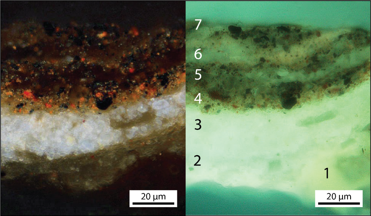
UV fluorescence image and visible light image (upper left and right) of a cross-section taken from the flesh shadows of Christ's right hand, showing an example from just one location of the complex pigment stratigraphy used by da Vinci to achieve sophisticated visual effects.
Layering (numbered in the image) includes the off-white imprimatura (4), predominantly composed of lead-white tinted by the addition of lead-tin yellow pigment, and complex layering (layers 4-7) composed of a lower brown-red layer dominated by vermilion and carbon black, followed by thin, semi-translucent pigmented glaze layers containing various mixtures of carbon and bone black with minor vermilion.
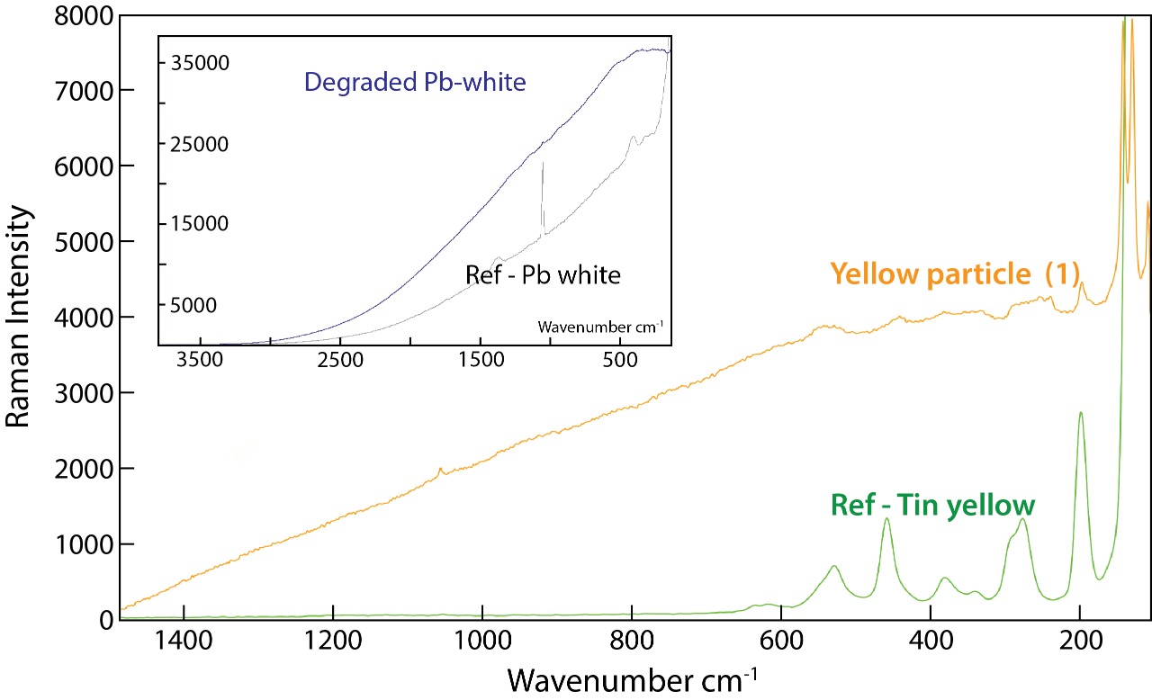
Compositions of pigments and their components are confirmed in the microRaman spectra shown here against reference materials for Pb-white and Sn-yellow. The yellow particle (number 1 in the upper image) shows the characteristic peaks for Sn-yellow. Measurements of white components suggest decay of the Pb-based pigment over time.
Learn More
Salvator Mundi: an investigation of the painting’s materials and techniques
Nica Gutman Rieppi, Beth A. Price, Ken Sutherland, Andrew P. Lins, Richard Newman, Peng Wang, Ting Wang, Thomas J. Tague Jr.
Heritage Science, 8:39, 2020