Virtual Tour for Pre-Clinical Imaging Solutions

Webinars
June 18, 11am EDT / 5pm CEST - Hyperpolarized xenon gas MR

Hyperpolarized 129Xe MR has been proven to be a powerful tool to evaluate the pulmonary function and has been widely used to study various lung diseases. Preclinical studies have advantages in exploring the feasibility of hyperpolarized 129Xe MR in diagnosing lung diseases and developing new techniques and application strategies.
Speaker: Dr. Xin Zhou, Vice director of Innovation Academy for Precision Measurement Science and Technology, Chinese Academy of Sciences (CAS)
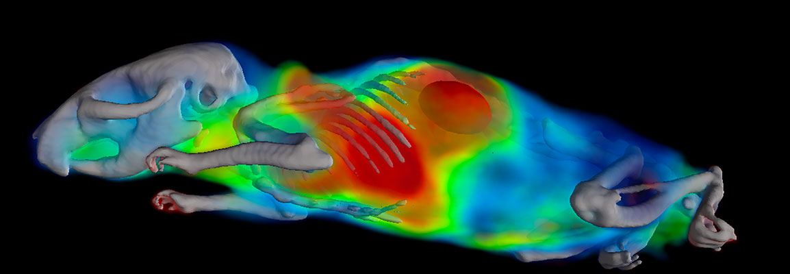
May 28, 11am EDT / 5pm CEST - microCT Applications in Lung Research and Immunology

The ability to quantitate lung disease and monitor disease development and resolution in real time is an advantage to using small animal microCT. In addition, 3D analysis provides data about the whole lung instead of the snap-shots typically associated with conventional analysis techniques, including histology. microCT provides the ability to assess a large diversity of lung diseases and the outcomes correlated to data generated from human patients.
Speaker: Dr. Elizabeth Redente, Associate Professor National Jewish Health
May 21, 11am EDT / 5pm CEST - PET Image Reconstruction Overview - Current and Future Trends

Image reconstruction is a fundamental part of PET allowing to generate 3D tomographic images of the tracer’s spatial distribution based on the position and timing of the detected annihilation gammas. PET reconstruction, common to other imaging modalities like CT and SPECT, apply algorithms whose mathematical details the general users may not need to understand. Nevertheless, a good knowledge on the performance characteristics of different algorithms can be highly beneficial to obtain reliable quantitative images more efficiently.
Speaker: Dr. Charalampos (Harry) Tsoumpas, Lecturer of Medical Imaging at the University of Leeds and Adjunct Professor at the Icahn School of Medicine at Mount Sinai, New York - Dr. Josep Oliver, Bruker senior NMI image reconstruction expert
May 7 - Estudos Pré-clínicos por Imagens – Mais uma Ferramenta no Processo de Pesquisa Farmacêutica
Assim que uma molécula candidata a medicamento é identificada e testada para sua eficácia inicial in vitro, o próximo passo é a realização de testes de eficácia e de segurança em animais, normalmente utilizando-se roedores, tais como camundongos e ratos.
Speaker:
Dr. Alan Nunes, Preclinical Imaging Product Manager Nova Analitica Importação e Exportação LTDA - Dr. Scott Ireland, Sales Manager, Preclinical Imaging Division
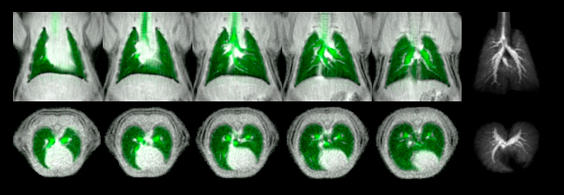
May 7 - Hyperpolarized 129Xe MRI of Pulmonary Gas-Exchange in Rodents

Hyperpolarized 129Xe MRI has emerged as a powerful technique to evaluate lung function and has found clinical application in several disorders like asthma, chronic obstructive pulmonary disease (COPD), idiopathic pulmonary fibrosis (IPF) and more. Preclinical 129Xe MRI has the potential to find new applications and accelerate clinical research, but is challenging and limited to only a few centers worldwide.
Speaker: Dr. Rohan Virgincar, MRI Applications Scientist at Bruker USA - Dr. Kristin Granlund, MRI Applications Scientist at Bruker USA
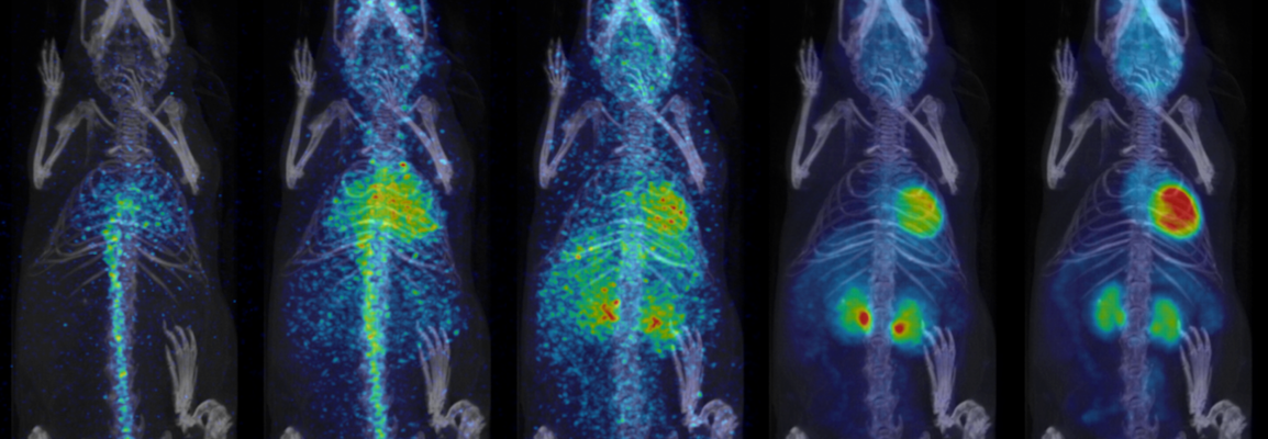
April 22 - Ensuring Quantitative Accuracy in PET
Positron emission tomography (PET) is not only a highly sensitive in vivo imaging technique, with a broad library of tracers, but it is inherently quantitative. This is the basis of the widely known metric SUV (Standard Uptake Value). However, producing accurate quantitative data requires the manufacturer to supply adequate tools and quality control protocols ideally in a user-friendly workflow. In addition, the image format must then maintain this accuracy and metadata necessary for SUV and tracer kinetic analysis, and image analysis software must utilize this information properly. Although it trails PET, advances in SPECT reconstruction and corrections have also brought quantitation to this modality.
Speaker: Cesar Molinos, Physicist and Electronics Engineer, Bruker BioSpin - Geoff Warnock, Senior Application Scientist, PMOD
March 5 - Ex Vivo MicroangioCT: Advances in Microvascular Imaging
Over recent years, non-destructive 3D imaging techniques such as micro-computed tomography (microCT) have increasingly captured the attention of preclinical researchers across almost every discipline. Now, a technique called microangioCT imaging is being used with a novel polymerizing contrast agent that enables 3D visualization of organ vasculature at unprecedented levels.
Speaker: Kjell Laperre, PhD, icro-CT Market Product & Applications Manager in the Bruker Biospin Pre-Clinical Imaging Division - Dr. Ruslan Hlushchuk, Lecturer and Group Leader at the Department of Topographic and Clinical Anatomy at the Universität Bern, Institute of Anatomy