Live Imaging in Cell and Developmental Biology
Life is on in 3D - just reveal it in real time!
Live-cell imaging is currently an integral part of modern developmental and cell biology. It enables the precise tracking of highly complex and dynamic molecular and cellular events during embryonic development and in cell culture applications. Light-sheet microscopy is the most powerful method to unveil the complex structure and behavior of whole embryos and cell cultures. It enables collecting meaningful live-cell imaging microscopy data by providing fast, long-term and high-resolution imaging while minimizing phototoxic effects and keeping environmental conditions stable.
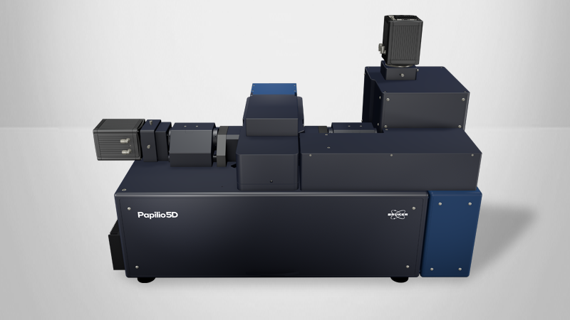
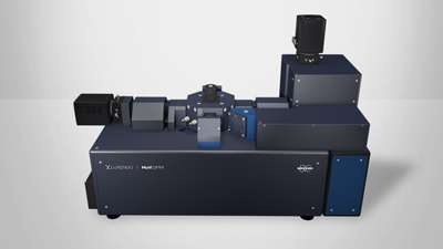
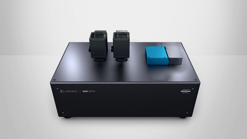
Bruker offers different light-sheet geometries for long-term rapid and gentle 3D imaging of living samples. The unique 4-axis concept of the Luxendo MuVi SPIM is the ideal tool for in toto imaging of Drosophila, Zebrafish, and many more sample types. The Luxendo InVi SPIM light-sheet microscopes are designed for live-sample imaging applications ranging from 2D and 3D cell cultures to small embryos, under precisely controlled environmental conditions. Expand your capabilities with the Luxendo InVi SPIM Lattice Pro and its flexible choice of illumination patterns – Gaussian beams, Bessel beams, optical lattices, and more.
Enjoy the flexibility and versatility of Luxendo SPIM solutions in terms of photo-ablation, photo-activation, perfusion etc.
Cell Tracking in Drosophila Embryo Development
Tumorigenesis in Mammary Organoids
Tracking Mitosis in HeLa Cell Culture
Luxendo Light-Sheet Microscopy Systems for Developmental and Cell Biology Applications
Do you want to image delicate cell cultures or whole living model organisms as they develop and grow? Start observing them in their native environment in 3D over days! Our light-sheet technology is the gentlest way to track these dynamic processes over long periods of time! Our choice of light-sheet microscopy systems enable high-resolution, dual sided-illumination imaging of delicate samples in their native, stable, controlled 3D environment.