

Bruker at Neuroscience
Come visit us in Booth 424!
October 5 - 9 | McCormick Place Convention Center | Chicago, IL, USA
Meet us in Chicago for the annual Society for Neuroscience conference, where we’ll be showcasing our suite of fluorescence microscopes and spatial biology solutions for life science imaging and analysis. Our solutions help scientists make breakthrough post-genomic discoveries and develop new applications that improve the quality of human life.
Connect with Bruker experts on the expo floor, at vendor talks, or at our annual symposium to hear case studies, learn about our technologies, and ask your questions. Our team is excited to help you discover how Bruker’s advanced solutions can help you see the biology of life more clearly! See you there!
Conference Host:
Society for Neuroscience
Conference Venue:
McCormick Place Convention Center | Chicago, IL, USA
Bruker’s Satellite Event on Saturday, Oct 5th
Multimodal Neuronal-imaging: From Synapses to Circuits
Satisfy your sweet tooth while expanding your mind at Bruker’s 6th annual symposium on multimodal fluorescence imaging for neuroscience. Hear from leaders in the field who are using cutting‑edge techniques, including one‑photon, two‑photon, super‑resolution, and light‑sheet—all while enjoying dessert on us. Don’t miss this exciting opportunity to dive into groundbreaking discoveries and advancements in neuroimaging!
This event is open to all SfN 2024 registrants.
Featured Bruker Talks at the Show
Simultaneous Multi-region Neuronal-Level Brain Imaging in Freely Behaving Mice
*T. MAHMOUDI1, R. CHRISTO2, Y. LEE2, C. SANTOS2, J. KANEM1, A. PAPANICOLAOU3;
1Bruker, Toronto, ON, Canada; 2Bruker LTD., Toronto, ON, Canada; 3Bruker LTD, Toronto, ON, Canada
Session Number: PSTR491
Session Title: Cutting-Edge Methods for Neuronal Monitoring and Control in Freely Moving Animals
Session Day and Time: 10/9/2024 1:00:00 PM-5:00:00 PM
Imaging and stimulating neurons in freely behaving rodents using a multi-color multi-region miniscope
A. PAPANICOLAOU, J. KANEM, T. MAHMOUDI, Y. LEE, R. CHRISTO, C. SANTOS, *Y. SOUDAGAR;
Bruker, Toronto, ON, Canada
Session Number: PSTR430
Session Title: Optogenetic Tools
Session Day and Time: 10/9/2024 8:00:00 AM-12:00:00 PM
Miniscope-based in vivo blood vessel imaging enables a novel assay for testing migraine therapeutic candidates
*J. ZAPATA1, K. ZITELLI1, J. J. NASSI1, B. J. HALL2, P. BOTTA2
1Inscopix Discovery Lab., Inscopix Inc., Mountain View, CA; 2Circuit Biol., H Lundbeck, Valby, Denmark
Session Number: PSTR067.10 / C108
Session Title: Headache, Migraine, and Trigeminal Circuits
Session Day and Time: 10/6/2024 09:00-10:00 AM
A circuit-based, neurobehavioral assay for preclinical antidepressant profiling
*J. NASSI, L. BELLIER, N. REN, S. HUANG, D. CHENG, L. GRAY, O. MILLER;
Inscopix Discovery Lab., Inscopix, Inc., Mountain View, CA
Session Number: PSTR182.07 / M31
Session Title: Major Depressive and Bipolar Disorder: Targeting and Treatment
Session Day and Time: 10/7/2024 10:00-11:00 AM
Preclinical neurobehavioral profiling of antipsychotics through miniscope calcium imaging
*S. HUANG, D. CHENG, L. BELLIER, O. H. MILLER, J. J. NASSI; Inscopix, Inc, Mountain View, CA
Session Number: PSTR375.07 / Z23
Session Title: Biomarkers and Therapeutics in Affective Disorders and Schizophrenia
Session Day and Time: 10/8/2024 3:00-4:00 PM
Highly multiplexed immunofluorescence imaging of human and murine brain tissues
X.MESHIK, M. INGALLS, A. NORTHCUTT, *O. BRAUBACH; Canopy Biosci. - A Bruker Co., St. Louis, MO
Session Number: LBA009
Session Title: Theme I Late-Breaking Posters
Session Day and Time: 10/8/2024 1:00-5:00 PM
Product Theaters:
Connect, Scale, Explore! Your New Inscopix Miniscope Ecosystem
Speaker: Brice Tiret, PhD
Date & Time: Sunday, October 6, 11:30 a.m.–noon
Location: MCP Exhibit Hall Aisle 900
Join us into the realm of integrated neuroscience. In the ever-evolving field, one must position themselves for success with new skills, technologies, and ideas. We present the latest multi-modal miniscope technologies, designed to enhance productivity and provide a unified environment to accelerate you to publication. Whether you're a scientist, a lab group, or in biotech, choose and scale the right solution for your research, saving valuable time and effort and transform your discoveries!
Imaging the Dynamics of the Nervous System
Speaker: Kevin Mann, PhD
Date & Time: Sunday, October 6, 1–1:30 p.m.
Location: MCP Exhibit Hall Aisle 900
Two-photon microscopy in neuroscience has unraveled many mysteries of perception, computation, and connectivity. Ca2+ imaging has illuminated brain function with resolution and precision at the scale of small networks, single cells, dendrites, and individual spines. Join us to learn how the Bruker Ultima 2Pplus and Investigator workstations combine holographic optogenetic activation, GRIN lenses, and the NeuraLeap rapid-focusing module to advance new frontiers in neural circuit interrogation.
Highest Plex Spatial Multiomics as a Discovery Tool for Cellular Aging and Pathogenesis in Alzheimer's Disease
Speaker: Alyssa Rosenbloom, PhD, Miranda Orr, PhD
Date & Time: Monday, October 7, 4–4:30 p.m.
Location: MCP Exhibit Hall Aisle 900
Alzheimer’s tau forms neurofibrillary tangles (NFT), correlated with neuron loss, cognitive decline, & cellular senescence. Exploring senescence transition, cell interactions, & tissue-wide effects requires high-resolution spatial solutions. CosMx SMI reveals spatially correlated gene modules & cell niches. High-plex multiomics, (GeoMx DSP), gives insight into NFT-associated senescence & neurotoxicity. A phospho-tau signature (CellScape) demonstrates rapid scale up model for disease evaluation.
Luxendo Light-Sheet and Acquifer Automated Microscopy: Smart 3D Imaging From High-Resolution to High-Throughput
Speaker: Malte Wachsmuth, PhD
Date & Time: Tuesday, October 8, 10–10:30 a.m.
Location: MCP Exhibit Hall Aisle 900
Experience 3D life across scales with Luxendo SPIMs for long-term imaging of living samples and the Acquifer IM for automated screening microscopy. Discover smart adaptive light-sheet fluorescence microscopy for long-term 3D imaging of both live, and large cleared biological samples. The Acquifer IM offers automated phenotypic screening with an intuitive imaging interface and the Acquifer HIVE is the complete solution for secure storage and processing of all imaging, sequencing, and omics data.
Bruker Satelite Event Abstracts
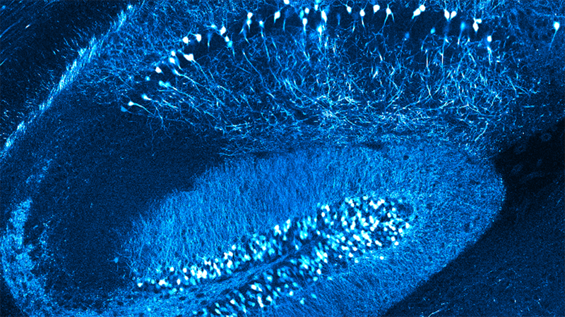
Illuminating cell-type-specific maps of the hippocampal spatial code
Author: Liset M. de la Prida, PhD, Research Professor
Affiliation: Instituto Cajal CSIC, Madrid, Spain
Abstract: Exploration drives the formation of hippocampal cognitive maps, yet understanding how they emerge from the activity of heterogenous cellular populations is unclear. We combine single- and dual-color imaging strategies to dissect cell-type specific contributions to the population code. Our data indicate that the hippocampus operates through parallel genetically-defined subcircuits to simultaneously encode local and global information from the environment in integrated cognitive maps.

The medial entorhinal cortex encodes multisensory spatial information
Author: Yi Gu, Investigator
Affiliation: National Institute of Neurological Disorders and Stroke
Abstract: Animals employ spatial information in multisensory modalities to navigate their natural environments. However, it is unclear whether the brain encodes such information in separate cognitive maps or integrates all into a single, universal map. We addressed this question in the microcircuit of the medial entorhinal cortex (MEC), a cognitive map of space. Using cellular-resolution calcium imaging, we examined the MEC of mice navigating virtual reality tracks, where visual and auditory cues provided comparable spatial information. We uncovered two cell types: “unimodality cells” and “multimodality cells”. The unimodality cells specifically represent either auditory or visual spatial information. They are anatomically intermingled and maintain sensory preferences across multiple tracks and behavioral states. The multimodality cells respond to both sensory modalities with their responses shaped differentially by auditory and visual information. Thus, the MEC enables accurate spatial encoding during multisensory navigation by computing spatial information in different sensory modalities and generating distinct maps.

Distinct timescales of recurrent dynamics in frontal and visual cortices
Author: Lucas Pinto, Assistant Professor
Affiliation: Department of Neuroscience, Northwestern University
Abstract: Cortical areas participate differently in cognitive computation. For example, experiments from my lab show that, during working memory, frontal areas get progressively more active with longer delays, while visual areas are only recruited during short delays. But what microcircuit mechanisms explain this duration-dependent recruitment of cortical regions? In this talk, I will discuss unpublished work from my lab aimed at answering this question. We use simultaneous targeted two-photon optogenetics and cellular-resolution calcium imaging to compare how the premotor (frontal) and primary visual cortices of the mouse sustain brief, focal excitatory input to the networks. I will show that frontal circuits are much more likely to respond to photostimulation, and additionally sustain very focal input much longer than visual circuits, by recruiting local activity sequences. Our findings suggest that stronger recurrent excitation in the frontal cortex may in part underlie longer activity timescales during spontaneous and cognitive behaviors.

Highest Plex Spatial Multiomics as a Discovery Tool for Cellular Aging and Pathogenesis in Alzheimer's Disease
Author: Miranda E. Orr, Ph.D.
Affiliation: Associate Professor Wake Forest School of Medicine Research Health Scientist Salisbury VA Medical Center
Abstract: Alzheimer’s tau forms neurofibrillary tangles (NFT), correlated with neuron loss, cognitive decline, & cellular senescence. Exploring senescence transition, cell interactions, & tissue-wide effects requires high-resolution spatial solutions. CosMx SMI reveals spatially correlated gene modules & cell niches. High-plex multiomics, (GeoMx DSP), gives insight into NFT-associated senescence & neurotoxicity. A phospho-tau signature (CellScape) demonstrates rapid scale up model for disease evaluation.

TBD
Author: Michael Barresi, Ph.D.
Affiliation: Professor of Biological Sciences
Smith College
Abstract: TBD

TBD
Author: Ege T. Kavalali, Ph.D.
Affiliation: Professor and Chair
Vanderbilt University
Abstract: TBD


Thank you
Your request was successfully sent.
You can email us with more information at productinfo@bruker.com.
If you would like to connect with a Bruker representative immediately, here are our regional offices:
Asia: +65 6540 4388
China: +86 10 5833 3000
Europe: +33 172 86 61 00
Japan: +81 3 3523-6361
North America: +1 805-967-1400