Bruker at Society for Neuroscience 2025
Visit Us at Booth 2513
November 16–19 | San Diego Convention Center | San Diego, CA
Connect with Bruker experts and explore our full suite of fluorescence microscopy technologies and spatial biology solutions for life science imaging and analysis, including:
- Two-Photon (2P) Microscopes for deep tissue imaging
- Miniscopes and Multi-Region Miniscope Microscopy for in vivo neural circuit studies
- Light-Sheet Microscopes for high-speed, high-resolution 3D imaging
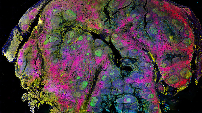
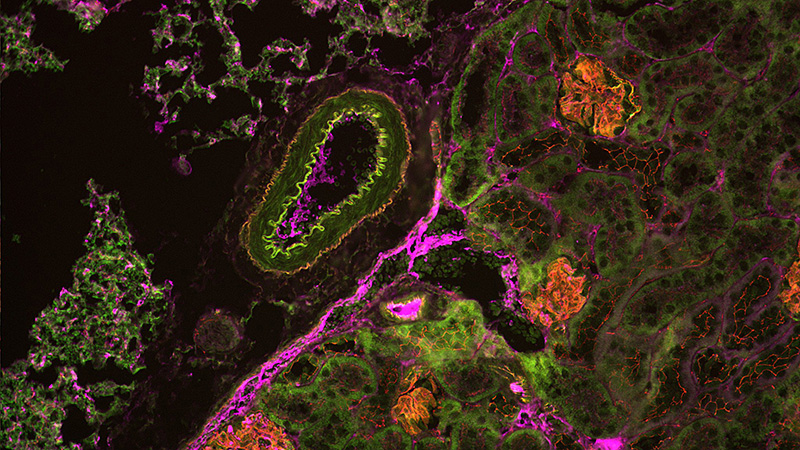
- Bruker Spatial Biology Solutions for high-plex spatial proteomics and transcriptomics
Our solutions help scientists make breakthrough post-genomic discoveries and develop new applications that improve the quality of human life. Our team will be available throughout the show to discuss applications, answer questions, and help you find the right solutions for your neuroscience research. See you there!
Conference Host:
Society for Neuroscience
Conference Venue:
San Diego Convention Center | San Diego, CA
Don’t Miss Our Presentations
We’re hosting several in-booth presentations and one Product Theater—stop by to hear case studies, technical insights, and application highlights from Bruker’s thought leaders. Full schedule coming soon!
Abstracts
In vivo longitudinal assessment of neural circuit dynamics and amyloid plaque progression in the motor cortex of an Alzheimer’s mouse model
Poster Number: PSTR111.07.
Date: November 16, 2025
Time: 1:00 PM - 5:00 PM
Location: SDCC Halls B-H
Acute and durable miniscope-based neurobehavioral profiling of 5-HT2A receptor agonists with hallucinogenic and non-hallucinogenic potential in mouse medial prefrontal cortex
Poster Number: PSTR355.06.
Date: November 18, 2025
Time: 1:00 PM - 5:00 PM
Location: SDCC Halls B-H
Pharmacologic activation of the muscarinic (M4) receptor modulates dorsal striatum neuronal activity before and after concurrent SSRI treatment
Poster Number: PSTR452.07
Date: November 19, 2025
Time: 1:00 PM - 5:00 PM
Location: SDCC Halls B-H
Product Theaters:
Illuminating Brain function through Two-Photon: Calcium and Voltage Dynamics with Optogenetics
Speaker: Jimmy Fong
Date: Sunday, November 16
Time: 4:00pm – 4:30pm
Location: SDCC Exhibit Hall Aisle 1200
Two-photon microscopy has advanced our understanding of computation and connectivity in neuroscience. Through in-vivo calcium imaging at cell-resolution, scientists study brain activity from scales of dendritic spines to neuronal networks. Combined with optogenetics and voltage imaging, 2P offers new insights into dynamic interactions in neural circuits. Learn how Bruker enables research with 3D holographic optogenetics, voltage imaging (OptoVolt), fast volumetric imaging (NeuraLeap), and more.
In-Booth Presentations
Unlimited Spatial Exploration in the Brain: Complete Biological Pathways-based multiomics with CosMx Whole Transcriptome
Speaker: Joe Beechem
Date: Sunday, November 16
Time: 10:00 AM - 10:30 AM
Illuminating the Brain: Mapping and Manipulating Neural activity in 3D
Speaker: Kevin Mann
Date: Sunday, November 16
Time: 10:30 AM - 11:00 AM
Abstract: Two-photon microscopy has been instrumental in neuroscience, revealing insights into perception, computation, and connectivity across diverse models. In particular, Ca2+ imaging has provided unprecedented resolution and precision at the scale of small networks, single cells, dendrites, and individual spines. Light-based manipulations, such as optogenetics, have enabled real-time circuit perturbations from single synapses to entire ensembles. I will discuss how Bruker’s advanced two-photon microscopy systems support modern imaging and stimulation experiments, offering high-speed acquisition, deep tissue penetration, and precise control for probing neural function.
nVista 2P: State of the Art Technology for Freely-behaving Animals
Speaker: Waylin Yu
Date: Sunday, November 16
Time: 11:00 AM - 11:30 AM
Abstract: The nVista 2P is a unique platform that brings high resolution optics to free-behavior neuroscience. It is an integrated, head-mounted system for two-photon imaging, leveraging Inscopix's best workflow solutions, like ProView Integrated GRIN lenses and IDEAS analysis platform. This presentation will cover how a single miniature microscope can image both shallow and deep brain regions, enabling use of fluorescent sensors for a wide range of applications, to capture neuronal, astrocytic, dendritic, or blood-flow dynamics.
A No-Brainer: An Integrated GeoMx DSP, and CellScape Workflow for Comprehensive Spatial Proteomics in Neurodegenerative Disease
Speaker: Oliver Braubach
Date: Sunday, November 16
Time: 1:00 PM - 1:30 PM
Big Data, Big Teams, Great Results: The Acquifer Hive for Large Data Acquisition and for Collaboration and Training in Teams
Presenter: Malte Wachsmuth
Date: Sunday, November 16
Time: 1:30 PM - 2:00 PM
Abstract: In this presentation we will demonstrate how the Acquifer HIVE platform enables the acquisition, processing and storage of large data sets as obtained in light-sheet microscopy, high-content screening or omics methods. It also serves as a powerful platform for data sharing for collaborative projects as well as for training and education purposes.
Imaging Neurological Tissue at Sub-Synaptic Resolution
Speaker: Abraham Kohrman
Date: Sunday, November 16
Time: 2:00 PM - 2:30 PM
Abstract: Synapses are the basis of communication between neurons within the nervous system. The structure and composition of synapses determines the type, frequency, intensity and duration of signaling across the synapse. While synapses are critical in neurological transmission they are difficult to study in situ, due to their size and the optically scattering nature of neurological tissue. Traditional optical microscopy methods offer resolution of roughly 250 nanometers, which means a roughly 100nm synapse (a typical size for a synapse) will appear at 2.5x its true size and contain no internal structural information. The Bruker Vutara VXL is a single molecule localization microscope designed to image with very high spatial resolution, on the order of 20nm in XY and 50nm in Z, providing substantially sub-synaptic resolution. Uniquely, due to its patented biplane Z localization technique, the Vutara VXL can provide sub-synaptic resolution tens of microns into neurological tissue samples, and similarly on cultured neurons close to the coverslip. This increased resolution allows for studies of the distribution of proteins or other biomolecules within the synapse, enabling better understanding of the structure and function of the synapse.
nVista 2P: State of the Art Technology for Freely-behaving Animals
Speaker: Waylin Yu
Date: Monday, November 17
Time: 10:00 AM - 10:30 AM
Abstract: The nVista 2P is a unique platform that brings high resolution optics to free-behavior neuroscience. It is an integrated, head-mounted system for two-photon imaging, leveraging Inscopix's best workflow solutions, like ProView Integrated GRIN lenses and IDEAS analysis platform. This presentation will cover how a single miniature microscope can image both shallow and deep brain regions, enabling use of fluorescent sensors for a wide range of applications, to capture neuronal, astrocytic, dendritic, or blood-flow dynamics.
Seeing Deeper, Seeing Better: Unveil the Next Dimension of Inverted Multiview Light-sheet Microscopy
Speaker: Malte Wachsmuth
Date: Monday, November 17
Time: 10:30 AM - 11:00 AM
Abstract: Join us for exploring how inverted light-sheet microscopy enables research on organoids and 3D cell cultures, ranging from high-resolution imaging e.g. of early mitotic events in mouse oocytes to investigating the behaviour of larger organoids under different conditions.
A No-Brainer: An Integrated GeoMx DSP, and CellScape Workflow for Comprehensive Spatial Proteomics in Neurodegenerative Disease.
Speaker: Oliver Braubach
Date: Monday, November 17
Time: 11:00 AM - 11:30 AM
Single Particle Tracking for Neuroscience Discovery
Speaker: Abraham Kohrman
Date: Monday, November 17
Time: 1:00 PM - 1:30 PM
Abstract: Neurons are large, dynamic cells. Active transport and compartmentalization are required to segregate and transport materials through the axon, dendrite and soma. Single particle tracking studies allow for the measurement of motion kinetics at very high resolution. Single particle tracking is the localization, and subsequent tracking of individual molecules or particles as they move within or between cells. Single particle tracking, as a super resolution technique provides information about the precise activity and behavior of biomolecules in living cells, otherwise unobservable through traditional microscopy techniques. Tracking particles in neurons could determine if the transport of materials down is active or diffusive, if structures in the cell membrane limit the diffusion of cell receptors, or if different cellular compartments or organelles have functional connections or contacts. Traditional 2d single particle tracks may not accurately measure particle motion dynamics, as biological materials do not constrain particles into a single plane. The Bruker Vutara VXL offers intrinsically 3d single particle tracking performance due to its patented biplane localization technique. Every particle within a roughly 1-micron volume can be localized and tracked, with high precision due to our high power, highly even illumination pattern and high quantum efficiency camera systems. 3d Single particle tracking of neurons could reveal new insights into the structure, function, regulation and control of the nervous system.
From Basic to Translational Research: The Universe of 1p Imaging
Speaker: Jonathan Nassi
Date: Monday, November 17
Time: 1:30 PM - 2:00 PM
Abstract: Bruker’s Inscopix miniscopes portfolio offers a unique platform to image, stimulate, and correlate various brain signals to behavior. This presentation will show how micro-environment affects neuronal activity in Alzheimer's research, and how metrics like blood flow, neuronal activity, and behavior correlate can be used in drug profiling and development.
From Basic to Translational Research: The Universe of 1p Imaging
Speaker: Jonathan Nassi
Date: Tuesday, November 18
Time: 10:00 AM - 10:30 AM
Abstract: Bruker’s Inscopix miniscopes portfolio offers a unique platform to image, stimulate, and correlate various brain signals to behavior. This presentation will show how micro-environment affects neuronal activity in Alzheimer's research, and how metrics like blood flow, neuronal activity, and behavior correlate can be used in drug profiling and development.
Innovative imaging solutions with Bruker: High speed sampling and imaging with Optovolt and xCore
Speaker: Kevin Mann
Date: Tuesday, November 18
Time: 10:30 AM - 11:00 AM
Abstract: Fast, high-SNR imaging is essential for resolving neural activity at millisecond timescales. Bruker’s OptoVolt scan-multiplying module enables kilohertz-rate voltage imaging over square fields of view, while maintaining compatibility with wide-field resonant scanning for calcium imaging. Combined with NeuraLight 3D patterned photostimulation, the system supports flexible experiments that integrate the imaging and manipulation of neural circuits. Bruker’s new xCore signal acquisition hardware further boosts performance with a custom data processing pipeline for precise PMT sampling and improved SNR. I will discuss how these tools expand the reach of multiphoton microscopy for fast, low-signal imaging in modern neuroscience.
Seeing Deeper, Seeing Better: Unveil the Next Dimension of Inverted Multiview Light-sheet Microscopy
Presenter: Malte Wachsmuth
Date: Tuesday, November 18
Time: 11:00 AM - 11:30 AM
Abstract: Join us for exploring how inverted light-sheet microscopy enables research on organoids and 3D cell cultures, ranging from high-resolution imaging e.g. of early mitotic events in mouse oocytes to investigating the behaviour of larger organoids under different conditions.
Big Data, Big Teams, Great Results: The Acquifer Hive for Large Data Acquisition and for Collaboration and Training in Teams
Presenter: Malte Wachsmuth
Date: Tuesday, November 18
Time: 1:00 PM - 1:30 PM
Abstract: In this presentation we will demonstrate how the Acquifer HIVE platform enables the acquisition, processing and storage of large data sets as obtained in light-sheet microscopy, high-content screening or omics methods. It also serves as a powerful platform for data sharing for collaborative projects as well as for training and education purposes.
Unlimited Spatial Exploration in the Brain: Complete Biological Pathways-based multiomics with CosMx Whole Transcriptome
Speaker: Joe Beechem
Date: Tuesday, November 18
Time: 1:30 PM - 2:00 PM
Bruker’s Satellite Event on Saturday, Nov 15
Registration for this event is now closed.
Exploring the Brain: All-Optical Investigation of Fast Neural Dynamics
Date: Saturday, November 15
Time: 7:00pm – 10:00pm
Location: Marriott Marquis San Diego Marina – Pacific Ballroom
Kick off SfN 2025 with Bruker’s 7th Annual Neuroscience Symposium, where leading researchers will share their latest discoveries using cutting-edge, all-optical technologies from Bruker. Hear from experts in the field, connect with your peers, and engage with the Bruker team about new advancements in neuroimaging. Don’t miss this chance to celebrate great science!
This event is open to all SFN 2025 registrants. Doors open at 7PM and presentations begin at 7:30PM. Reception to follow. Spots are limited, so reserve yours today!
Satellite Event Talks
Abstract: Animals rely on affordances, the meaningful possibilities for action offered by the environment, to navigate and interact with the world. However, how such affordance information arises in the brain remains unclear. Using in vivo one- and two-photon calcium imaging in freely moving conditions, we found that neurons in the anterior cingulate cortex (ACC) abstract spatial geometry into action-relevant representations. The ACC exhibits a hierarchical categorization of spatial geometry, including boundary, intersection boundary, and connectivity (referred to as spatial concept cells), and about half of these cells show self-object egocentric tuning. In particular, these neurons display different egocentric directions depending on the animal’s behavioral needs in the environment. The activity of these spatial concept cells and their egocentric tuning are maintained across days and across distinct environments. In summary, the ACC integrates abstract representations of spatial geometry with egocentric information to create the meaning of space, which leads to possible choices for action.

Abstract: Animals must execute appropriate actions for survival. In the brain, the striatum is a critical center for movement and learning and striatal activity correlates with vigor and kinematics of overt movements. However, the exact details of striatum control are less clear. A classical view has been that the striatum controls when and how vigorously to move through two opponent pathways. Specifically, the two populations of spiny projection neurons (D1-SPNs heading the striato-nigral pathway and D2-SPNs heading the striato-pallidal pathway) are thought to have opposing effects on movement. Other work indicates D1- and D2-SPNs may control specific actions in concert. Here, we performed 2-photon imaging and stimulation in the dorsolateral striatum as mice performed two forelimb actions in a self-paced manner, consisting of a push or pull isometric force on an immobile joystick. Both D1- and D2-SPNs populations equally predicted the preparation and execution of specific actions, irrespective of what action was reinforced. Further, we developed a closed-loop system to model and manipulate action-specific neural ensembles using holographic optogenetics through a GRIN lens. Stimulation of action-specific ensembles of both D1- and D2-SPNs increased the force of action, but only for their congruent action. These results show that D1- and D2-SPNs can control specific ongoing actions concurrently, with specific ensembles controlling actions as granular as forces exerted by the same body part.

Abstract: Understanding how neural circuits perform computations and store information is crucial for understanding the brain. This requires that we can read and write activity patterns in genetically defined neurons at cellular resolution and with millisecond precision during behaviour. I will describe experiments in which we have used Bruker microscopes to implement an “all-optical” strategy for interrogating neural circuits which combines simultaneous two-photon imaging and two-photon optogenetics. This strategy allows the physiological patterns of network activity to be read out, reproduced and manipulated in real time, enabling closed-loop feedback control of activity. I will discuss how these approaches can be used to trigger and read out synaptic plasticity in neural circuits, understand the dendritic basis of communication between neighbouring brain areas, and make new causal links between activity patterns in neural circuits and behavior.

Satellite Event Speakers

Takashi Kitamura, Associate Professor, Department of Psychiatry and Neuroscience, University of Texas Southwestern Medical Center
Darcy S. Peterka, Ph.D., Senior Scientist, Director of Team Science and Scientific Director of Cellular Imaging at Columbia's Zuckerman Institute

Michael Hausser, Director, (School of Biomedical Sciences), Faculty of Medicine; Chair Professor of Neuroscience; Lee Man-Chiu Professor of Neuroscience, The University of Hong Kong