
Element Mapping for Life Science
Element Distribution Analysis of Biological Specimens and Soft Materials in the Electron Microscope
The analysis of inorganic materials using energy dispersive X-ray spectroscopy (EDS) in electron microscopes is widely established, even in processes as routine as semiconductor fab lines and microscope-units along excavation belts in mining.
Mapping of the element distribution for solving problems in life science and medical research is however not so well known. Nevertheless, there is a huge potential for understanding biological specimens and soft materials nowadays, which is possible even down to atomic resolution and ppm level, offered by modern detector and microscope technology. Light elements, important for life processes, such as
- calcium, nitrogen, phosphorus and sulfur, can be routinely identified and their amounts quantified,
- even if the specimen is osmium stained.
Also, correlative approaches can secure the connection to light and X-ray microscopy and analysis.
We show EDX (EDS) data acquired using modern Bruker XFlash® technology and various electron microscope techniques including SEM and STEM to demonstrate the capabilities and value of element mapping and quantitative analysis in specimens relevant for biology, biomimetics, bio-mineralization and the interface of soft and hard materials. This includes the element distribution in cells, such as yeast or human blood cells infected with malaria, sulfur storing bacteria, nanotoxicity and the study of biomineralization. Furthermore, other analysis techniques available in electron microscopy and complementary to EDS and considerations for EDS in-situ, in liquid and in ice are discussed.
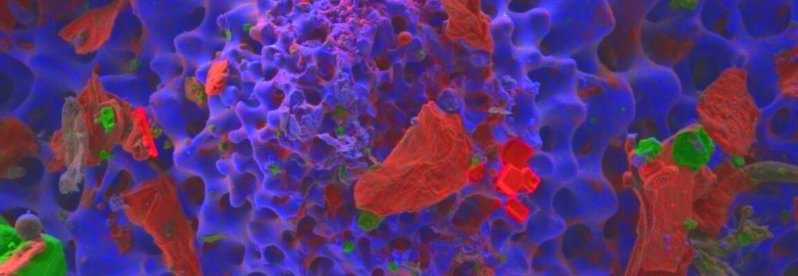
The figure depicts a spine tubercle of a sea urchin, which is the base of one of the long dangerous spines. The spine grows from there and is attached by collagen and a muscle ring, which allow movement and a lock and unlock mechanism (information source: California Academy of Science).
Who Should Attend
- Biologists interested in microscopy, micro- and nanostructure and the distribution of specific elements
- Students and professionals working at the interface of soft/organic and hard/inorganic matter
- Materials scientists
Speakers
Dr. Meiken Falke
Global Product Manager EDS/TEM, Bruker Nano Analytics
Max Patzschke
Application Scientist EDS, Bruker Nano Analytics
Watch this Webinar On-Demand
Please enter your details below to gain on-demand access to this webinar.
