The brain consumes approximately 20% of the body’s energy and receives 10-15% of blood from the heart. Since neurons have little reserve energy, they depend on a constant supply of glucose and oxygen from the blood. This is achieved by a vital process called functional hyperemia, which couples blood supply with energy demand, and requires signaling between synapses and the endothelial cells that make up the blood vessels in the brain.
Previous work by the authors showed that corticalarteriole lumen diameter is regulated by N-methyl-D-aspartate receptors (NMDARs) expressed by brain endothelial cells. The purpose of the current study was to determine whether endothelial NMDARs (eNMDARs) regulate functional hyperemia in vivo by comparing hemodynamic responses to whisker stimulation in awake eNMDAR loss-of-function mice with the responses of wild type mice.
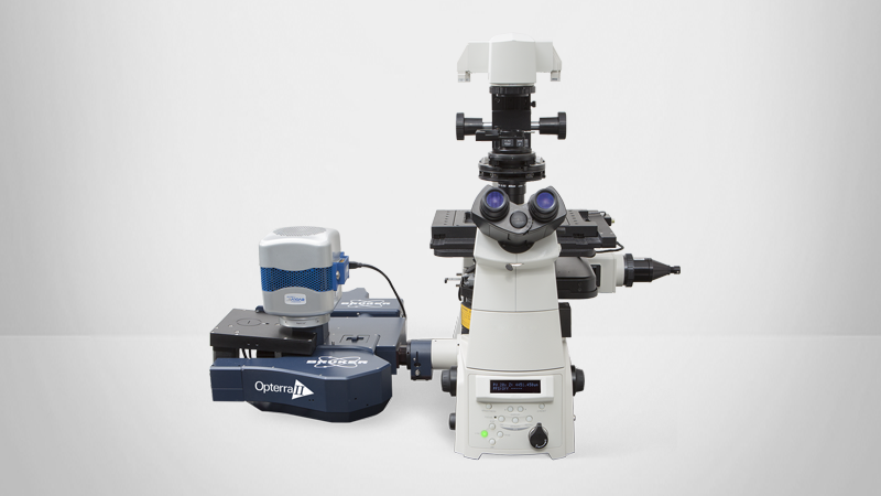
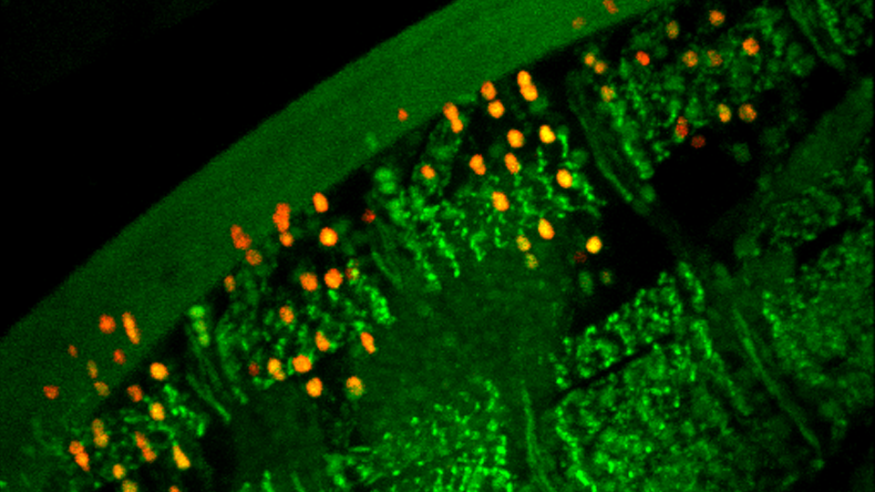
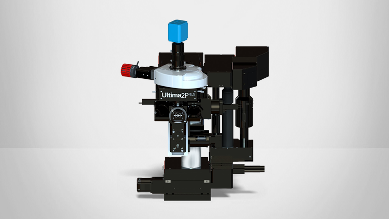
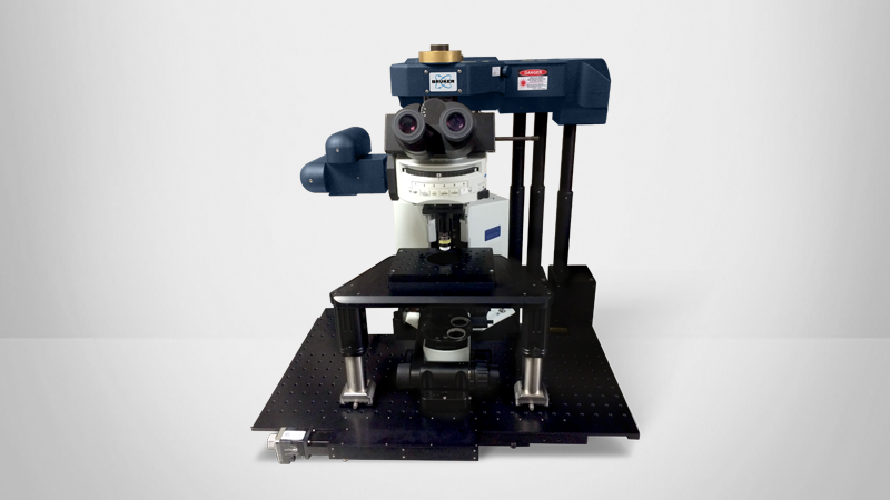
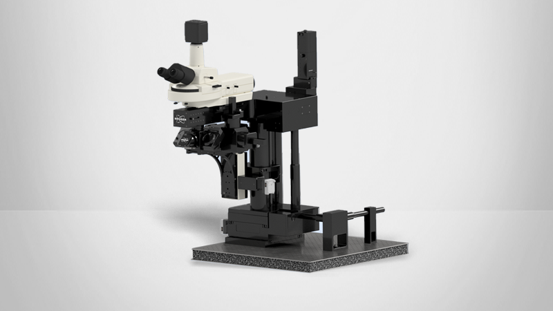
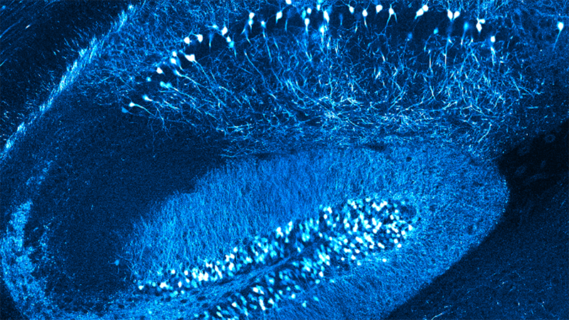
A Bruker Ultima IV multiphoton microscope was used to map the vascular network in the cortex to measure neuronal activity, and, using the line scan mode, to calculate red blood cell velocity, lumen diameter, and plasma flux following whisker stimulation in both living eNMDAR loss-of-function mice and wild type mice. Before imaging, the mice were injected with FITC-dextran to label plasma, and in some of the experiments, the mice received intracortical Fluo-4 (Ca2+-sensitive dye) and SR101 (astrocyte label) for monitoring neuronal activity. The stage of the Ultima IV multiphoton microscope afforded adequate room for an air-supported mobile cage, which allowed convenient experimental manipulations of the living mice.
In response to whisker stimulation, regional cerebral blood flow and hemodynamic responses were assessed in the barrel cortex of awake wild type or eNMDAR loss-of-function mice using multiphoton microscopy. Topological maps of vascular signals were created to a depth of ∼300 μm in layer II/III of the barrel cortex. Hyperemic enhancement of cerebral blood flow and vasodilation throughout the vascular network was observed in wild type mice, whereas in the eNMDAR loss-of- function mice, the hyperemic responses were reduced in both cerebellar blood flow and plasma flux in individual vessels.
The discovery of an endothelial receptor that regulates brain hyperemia in vivo provides insight into how neuronal activity couples with endothelial cells in the living brain. This study adds weight to the theory that the cerebral endotheliumis necessary for hyperemic cerebral blood flow regulation, and that diseases of endothelial dysfunction contribute to cognitive decline.