Languages
Receive future Journal Club updates via email.
This review appeared in the March 2022 edition of the Fluorescence Microscopy Journal Club — a monthly email brief highlighting leading-edge research and the latest discoveries supported by Bruker fluorescence microscopes.
Single-molecule localization microscopy (SMLM) is a powerful tool for visualizing and quantifying nanoscale organization across a variety of biological systems. However, the ability to image tissue samples in 3D and at high resolutions presents a challenge for many SMLM systems. This is partially due to limitations of instrumentation along with complex sample preparation requirement; standard total internal reflection (TIRF) systems restrict imaging to the coverslip surface and preparing thin tissue slices can be a daunting task.
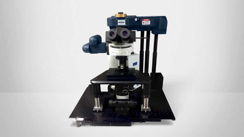
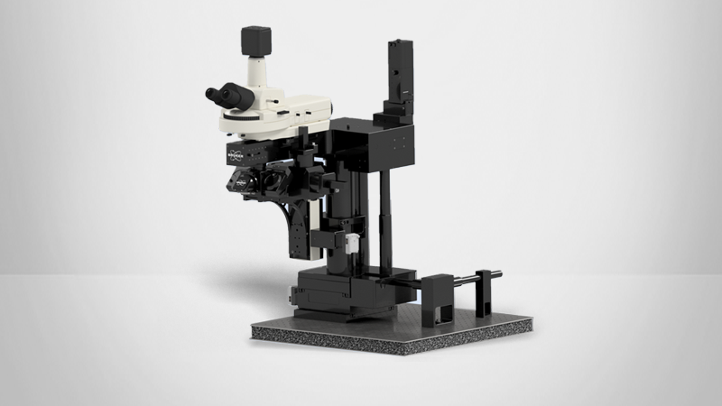
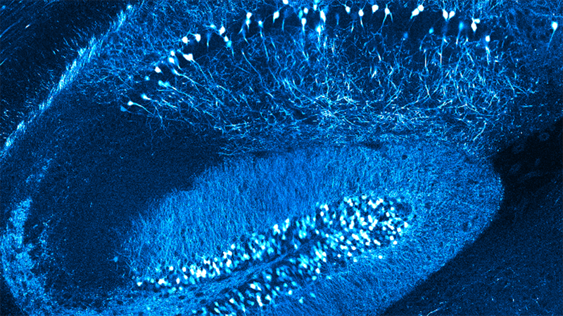
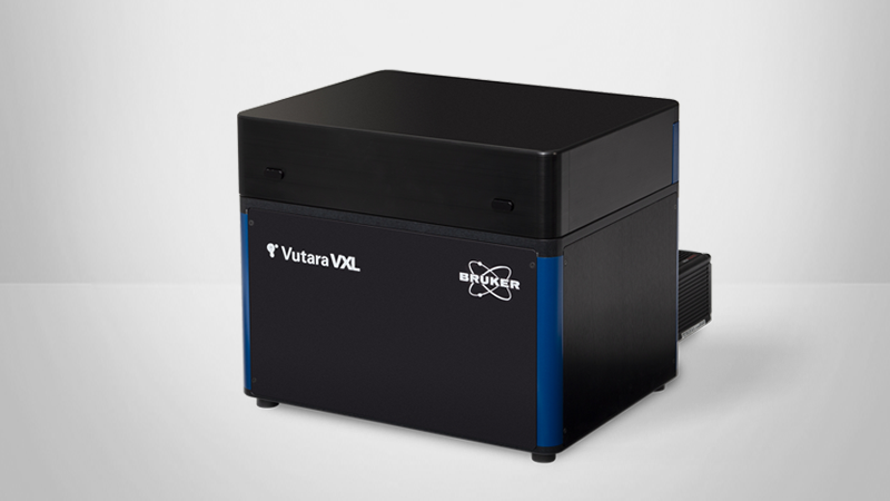
In this paper from Albrecht et al., researchers conducted a comprehensive experiment to optimize sample preparation conditions for 10µm-thick retinal tissue sections. Using Bruker’s Vutara 352 microscope and SRX software to execute a new super-resolution imaging method — RAIN-STORM (Rapid Imaging of Tissues at the Nanoscale Stochastic Optical Reconstruction Microscopy) — the authors:
In the case of these 10µm sections, the Vutara microscope is the ideal system for 3D imaging due to its epi-illumination and biplane detection means of obtaining 3D data with every acquisition without the need for an astigmatic lens. As a result, they were able to image the entirety of the 10µm sections by scanning in Z with more accuracy at larger depths than would be obtainable with astigmatic TIRF-based 3D imaging. For quantifying each image, researchers assessed the total number of localizations, sample detail preservation, background signal, and planar and axial resolutions. OPTICS (Ordering Points to Identify the Clustering Structure) and Fourier Ring Correlation analysis, which are available directly in the SRX statistical analysis suite, were used to identify associated contiguous structure from unstructured data, and to assess planar and axial resolution of all images.
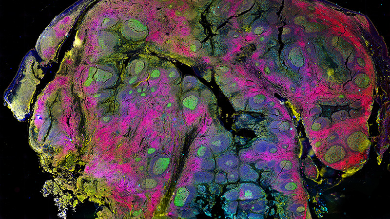
The optimized conditions were tested on and are applicable to a wide range of tissue types and many molecular targets and biological structures. Furthermore, the optimized protocol can be successfully applied to clinically derived human tissue.
KEY TERMS: