CellWizard Stage

Bruker's CellWizard™ Stage delivers pioneering, multi-compartment AFM imaging and nanomechanical analysis capabilities for life science applications, greatly extending the range, speed, and versatility of experiments possible, and pushing the boundaries of performance on the nanoscale. It facilitates automated large-scale mapping, high resolution imaging, and nanomechanical analysis in each chamber, and is ideal for the in vitro investigation of challenging biological samples.
Redefining the Future of BioAFM
The CellWizard Stage embodies state-of-the-art precision engineering. Using artificial intelligence (AI), it identifies sample features of interest and navigates autonomously from well to well, performing systematic, reproducible, and highly accurate measurements in each compartment. This next-generation AFM accessory delivers automated, multiscale, and multiparametric analyses. The ability to investigate biological samples in vitro, over a large scan range, and in a multi-compartment environment, allows a more comprehensive analysis and a deeper understanding of complex biological mechanisms, such as cellular communication and immune response.
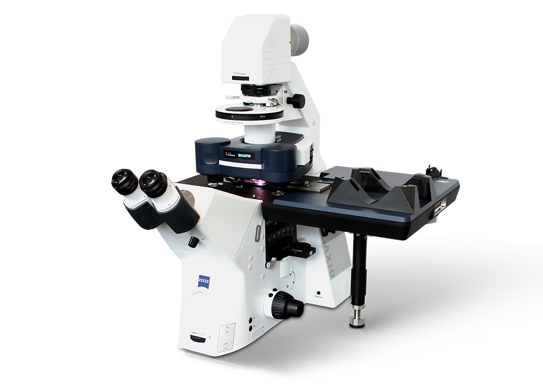
The CellWizard Stage is compatible with the Bruker BioAFM atomic force microscope platform and can be seamlessly integrated into advanced optical microscopes.
Superior Performance
- Compatibility with select choice of commercially available multiwell plates and multi-compartment glass slides
- Fast and precise positioning with state-of-the-art encoders and custom developed firmware
- Operation with renowned Bruker modes and features, including PeakForce Tapping and SmartMapping
- Innovative software packages, such as, the AI optical image-guided AutomaticNavigation feature and the extended DirectTiling feature that now includes optical stitching and background correction
New Possibilities
- Trigger different enzymatic reactions in each well
- Study cellular response to varying stimuli
- Deliver separate active ingredients to biological targets
- Study biofilm formation inhibition factors
- Quantify adhesion and disaggregation on varying substrates

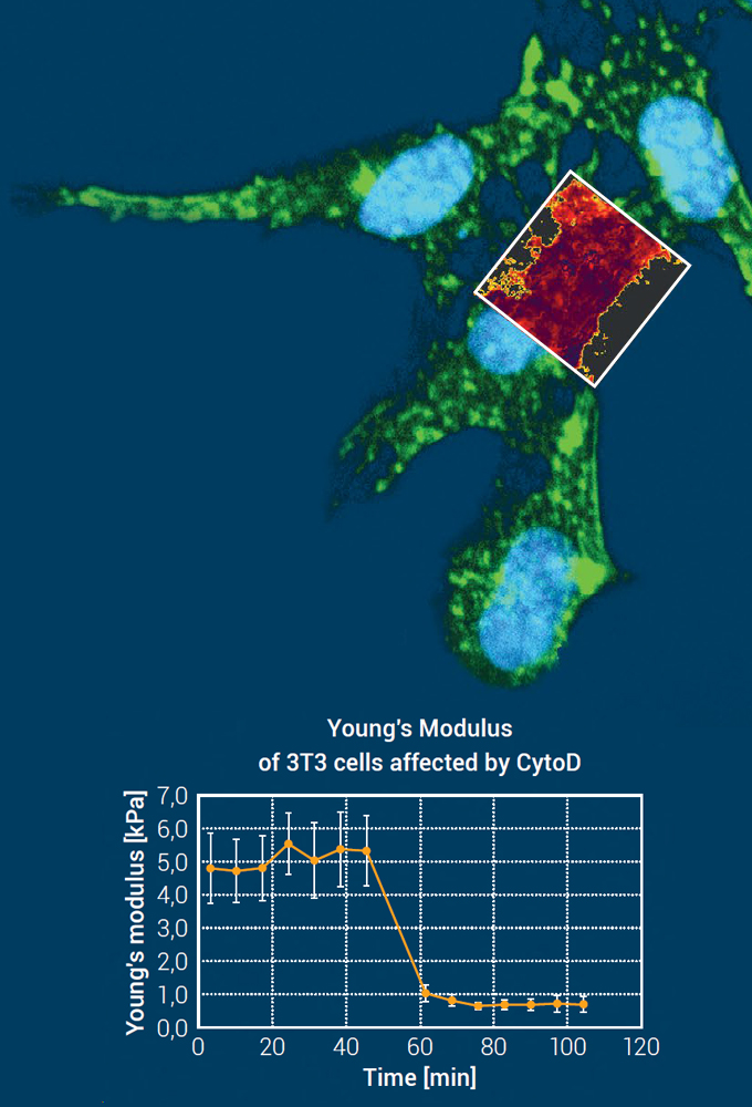
The time-dependent effect of cytochalasin D on the mechanical properties of 3T3 cells is shown in the graph below. The depolymerization of the actin filaments results in a reduction of the Young’s modulus over time.
QI scan size: 25 μm × 25 μm
Young’s modulus range: 5.0 kPa.
Sample courtesy of Dr Wedepohl, Freie Universität Berlin, Germany
Unmatched Functionality
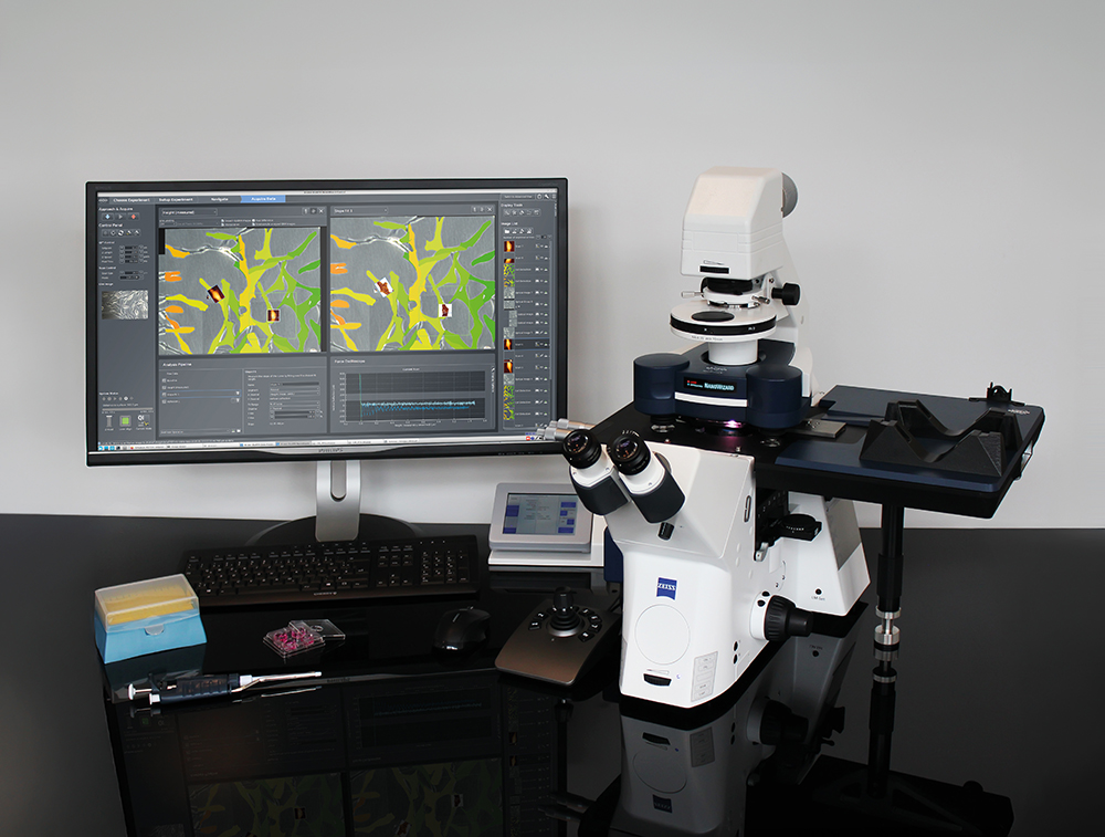
Outstanding Automation
CellWizard Stage encompasses state-of-the-art automation features and advanced machine-learning capabilities that significantly improve the accuracy and effectivity of repetitive workflows by alleviating user error and optimizing productivity.
The AI-guided optical segmentation tool uses optical images to identify sample features of interest and automatically creates MultiScan lists within or surrounding them.
Optical snapshots or scheduled rinsing steps can be added to the planned list, and the instrument will then run measurements autonomously, with the CellWizard Stage safely changing between compartments as specified.
The AI image segmentation can use either the pre-trained models provided or user-trained custom models tailored to individual applications.

Comprehensive Capabilities
Explore a wide spectrum of easy-to-use features and options, such as:
- Extensive optical integration options that include the DirectOverlay 2 software feature and accessories for operation on inverted optical microscopes
- Multi-chamber substrate supports:
4-well plate holder, a holder for four separate 35mm Petri dishes, and a holder for two standard glass slides - Bruker BioAFM PetriDishHeater options for 35mm and 50mm dishes
- SmartMapping option with free-hand ROI-selection and intelligent piezo and motor motion – ideal for challenging biological samples
- PeakForce QNM, QI Advanced mode, and microrheology options for quantitative nanomechanical analysis
- Hinge-clip cantilever holder for easy operation in confined chambers
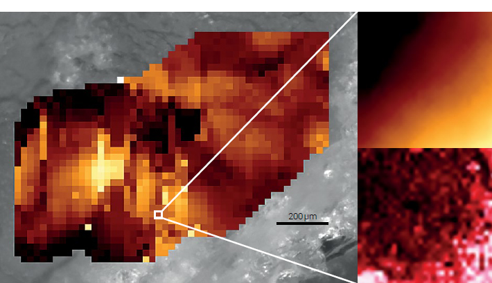
Corresponding AFM topography (upper right) and Young’s Modulus (lower right) of a zoomed-in scan: Scan size: 30 μm × 30 μm Pixel size: 1μm; Height range: 35 μm; Young’s modulus range: 8.0 kPa
Sample courtesy of Prof. Sack, Charité-Universitätsmedizin, Berlin, Germany
Extensive Flexibility
CellWizard Stage can be seamlessly combined with advanced optical techniques, such as confocal and super-resolution microscopy, for enhanced analysis capabilities and precise, correlated, multiparametric measurements.
It is compatible with the Bruker BioAFM family of instruments and its extensive range of modes, add-ons, and accessories, e.g.:
- Humidity control
- QR code reader for probes
- Nanolithography and Nanomanipulation
- ExperimentPlanner
- Cytosurge FluidFM® options
- Focus tracking support
The CellWizard Stage delivers the flexibility necessary for interdisciplinary research facilities and collaborative scientific environments.
Selected Relevant Publications
- Shioka et al., Ex vivo SIM-AFM measurements reveal the spatial correlation of stiffness and molecular distributions in 3D living tissue. Acta Biomat. (2024), 189, 351–365, https://doi.org/10.1016/j.actbio.2024.09.023
- Powell et al., Cisplatin resistance alters ovarian cancer spheroid formation and impacts peritoneal invasion, Front. Cell Dev. Biol. (2025)., https://doi.org/10.3389/fcell.2025.1450407
- Sunadome et al., Directionality of Developing Skeletal Muscles Is Set by Mechanical Forces. Nat Commun 2023, 14 (1), 3060, https://doi.org/10.1038/s41467-023-38647-7
- Brauer et al., Mechanical Heterogeneity in a Soft Biomaterial Niche Controls BMP2 Signaling. Biomaterials 2024, 309, 122614. https://doi.org/10.1016/j.biomaterials.2024.122614
- Villeneuve et al., Mechanical Forces across Compartments Coordinate Cell Shape and Fate Transitions to Generate Tissue Architecture. Nat Cell Biol 2024, 26 (2), 207–218.
https://doi.org/10.1038/s41556-023-01332-4
The Widest Range of Accessories on the Market
Optical systems/accessories, electrochemistry solutions, electrical sample characterization, environmental control options, software modules, temperature control, acoustic and vibration isolation solutions and more. Bruker provides you with the right accessories to control your sample conditions and to perform successful experiments.
Accessories to extend your BioAFM capabilities
Bruker offers an extensive range of BioAFM system add-ons, accessories, and modes to deliver maximum experimental and sample control, superior versatility, and enhanced useability. These options extend the range of applications and experiments supported by Bruker BioAFMs far beyond what is possible with any other BioAFM system on the market today.
Available options include optical systems/accessories, electrochemistry solutions, electrical sample characterization, environmental control options, software modules, temperature control, acoustic and vibration isolation solutions, and more.
Browse our online accessories database or download our accessories handbook to learn more.

Watch Recent BioAFM Webinars
Our webinars cover best practices, introduce new products, provide quick solutions to tricky questions, and offer ideas for new applications, modes, or techniques.
Testimonials

The CellWizard Stage is a milestone development - it introduces AI-guided approaches in AFM, significantly enhancing the speed and accuracy of stage dynamics, and providing multi-throughput measurements from precise, repeatable spatial locations. AFM is now more applicable than ever, collecting large-scale, high-resolution data sets, across a range of 3D cell samples (cells, tissues, spheroids and organoids) essential for cell and molecular biology analysis. The multiwell format allows us to incorporate biological variables and the full array of BioAFM measurements in a single experiment format.
Prof. Lewis Francis, Biomedical Sciences, Swansea University, UK