RAMANwalk
Measure Smarter.
Not Harder.
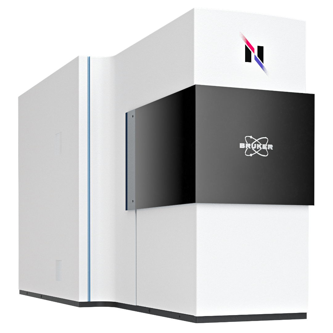
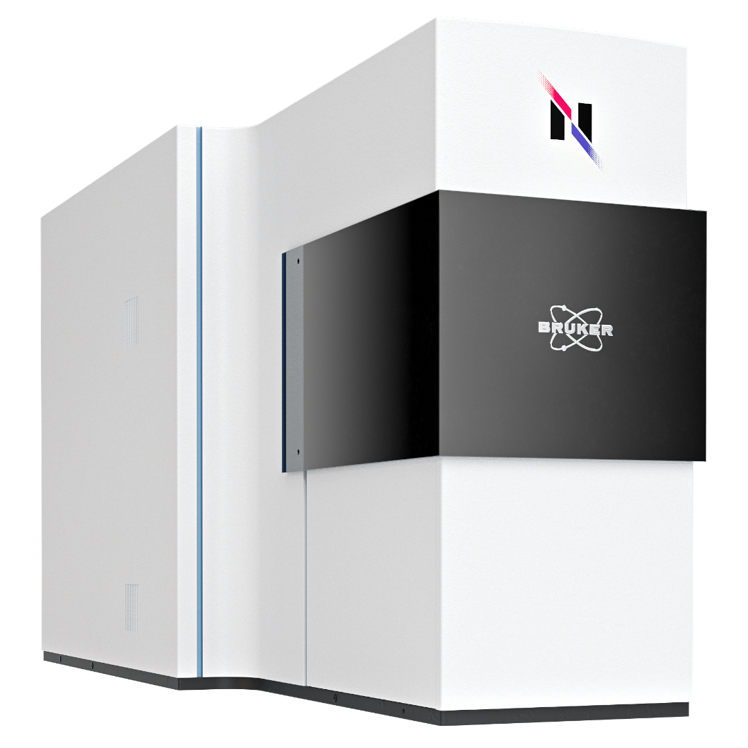
A Raman Microscope Driven by a More Intelligent Measurement Strategy.
Powered by stochastic sampling and high-speed galvano scanning, RAMANwalk continuously recognizes Raman patterns as they appear. The system automatically determines the most efficient measurement path, focusing on the regions that matter most and delivering Raman images more than five times faster than conventional scanning approaches.
Ease of use meets highest efficiency in Raman microscopy.
The RAMANwalk follows a new approach compared to traditional Raman microscopes - and that's why it stands out from the crowd. Highest spectral quality and super fast Raman image previews make it the perfect choice for routine.
The RAMANwalk offers:
- Reduce imaging time by random-walk technology
- Galvanic mirror for laser beam steering
- High spectral and spatial resolution
- Completely automated hardware
- Powerful analysis for 2D/3D Raman images
- Auto-calibration and auto-alignment
The Best RAMANwalk Features and How They Help You.
Comparing laser point scanning to "table" or raster scanning.
Conventional Raman microscopes typically follow a predefined raster pattern. RAMANwalk takes another route. Galvanic mirrors move the laser rapidly across the sample, while its Random Walk approach actively searches for Raman signals using chemical rather than purely visual information.
Once a Raman signal is identified, RAMANwalk investigates the surrounding area. When that signal disappears, the system reduces the probability assigned to the region and rapidly explores elsewhere. The result is a smarter route through the sample and exceptionally fast Raman preview images.

See meaningful Raman information sooner
RAMANwalk’s Random Scanning generates an early chemical preview by acquiring Raman spectra at distributed positions across the sample. These measured spectra provide the spectroscopic basis for estimating the information between sampled positions and constructing an interpolated Raman image.
As acquisition progresses, interpolated information is replaced by directly measured Raman data, allowing users to assess relevant chemical and spatial features before the complete measurement is available.
- Example 1: Distinguishing polymer beads
- Example 2: Locating a target mineral in ore
- Example 3: Mapping caffeine in a pharmaceutical tablet

Differentiate visually similar polymers
Polystyrene and PMMA beads are difficult to distinguish in the visual image. Raman imaging differentiates the polymers through their characteristic spectra and maps polystyrene in green and PMMA in purple. The interpolated image after 10 minutes already reproduces the main chemical and spatial features of the completed 23-minute measurement. This gives users an earlier overview of the sample while the acquisition continues.

Locate minerals of interest in complex ore samples
Raman spectroscopy identifies minerals through their characteristic vibrational spectra, providing information that complements elemental analysis. In this heterogeneous ore sample, the color-coded Raman image reveals the distribution and spatial associations of different mineral phases. The interpolated seven-minute image already indicates regions where the target mineral occurs, compared with the completed 20-minute measurement. This can support mineral exploration, ore characterization and decisions about further analysis.

Visualize ingredient distribution in pharmaceutical formulations
Raman imaging differentiates pharmaceutical ingredients through their characteristic spectra and maps their distribution within a formulation. In this tablet, caffeine is highlighted in red alongside other components shown in green and blue. The interpolated six-minute image already reveals the principal caffeine distribution, compared with the completed 30-minute measurement. Such chemical images can support formulation development and the assessment of ingredient distribution, domain size and agglomeration.


RAMANwalk specifications.
| Spatial Resolution | 350 nm in X and Y; 1 µm in Z | |
| Objective Lenses | 5x, 10x, 20x, 50x, 100x | |
| Spectral Resolution | <1.5 cm-1 (depends on grating, up to 3 gratings available) | |
| Stage Details | 30 * 30 * 35 mm XYZ-motorized stage | |
| Calibration | Auto-calibration based on standard lamp and sample | |
| Alignment | Auto-alignment of optical path | |
| Laser Safety | Laser safety class I door with interlock |