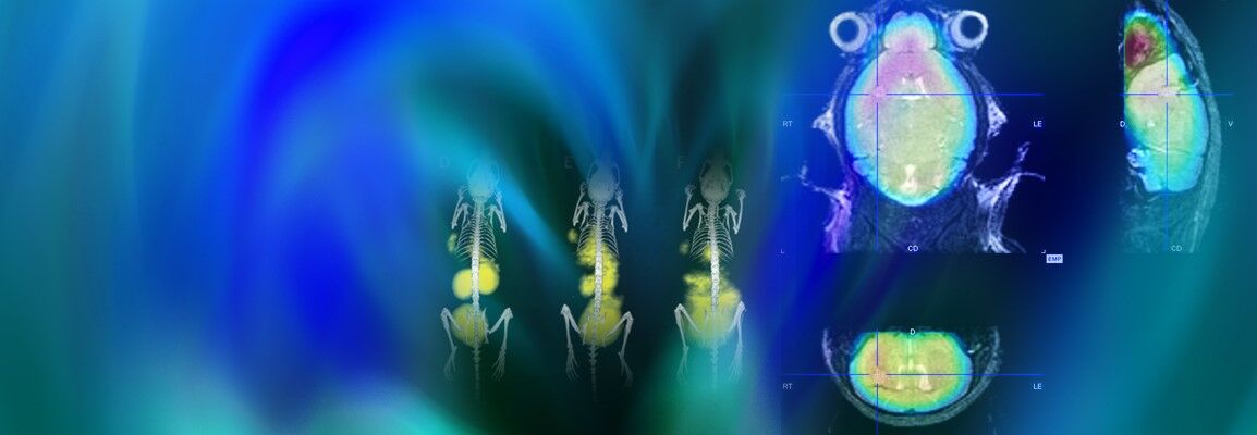
Advancing Cancer Research and Therapeutic Development with In Vivo Imaging
Cancer remains one of the world’s most significant healthcare challenges, driving efforts to develop more effective and personalized treatments. In the clinic, imaging supports cancer detection, diagnosis, staging, treatment planning, and monitoring of therapeutic response.
In preclinical oncology research, non-invasive in vivo imaging enables researchers to investigate tumor biology, disease progression, metastasis, treatment efficacy, and drug safety in real time within the complex environment of a living model.
Longitudinal Imaging for Preclinical Cancer Research
By combining complementary anatomical, functional, and molecular modalities, researchers can monitor changes longitudinally in the same animal. This approach also supports the reduction and refinement principles of the 3Rs by increasing the information obtained from each animal while reducing the number needed to achieve statistical relevance.
Imaging Technologies for Oncology Research
A wide range of imaging technologies contribute to oncology research, providing complementary insights into tumor biology, progression, and treatment response:
- Optical imaging, including BLI and FLI:
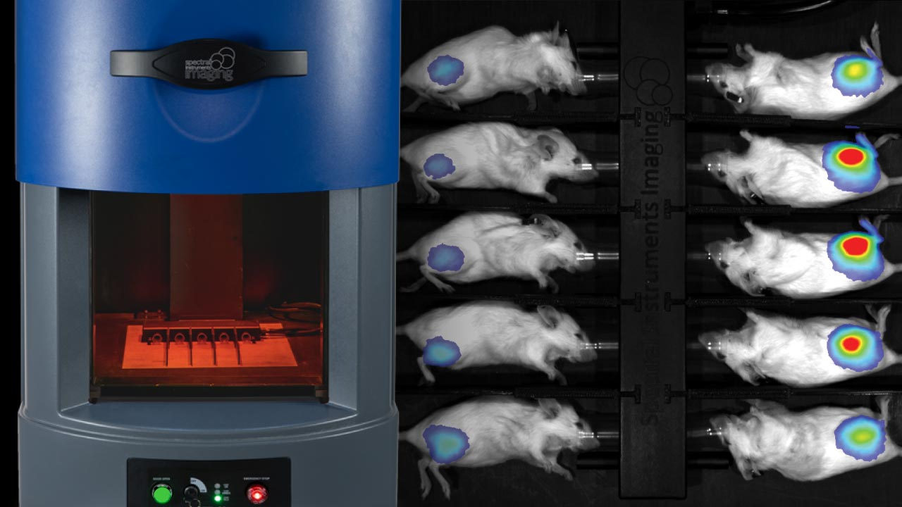
Bioluminescence imaging (BLI) and fluorescence imaging (FLI) enable highly sensitive detection of cellular and molecular processes in vivo. This approach allows for very early detection of tumor growth, including metastasis.
- MRI:
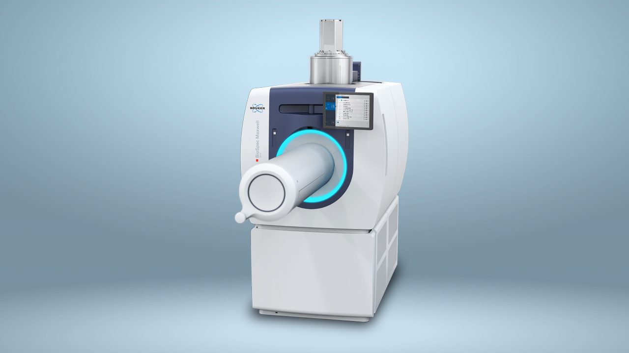
Delivers detailed soft tissue characterization and functional information.
- PET and SPECT:
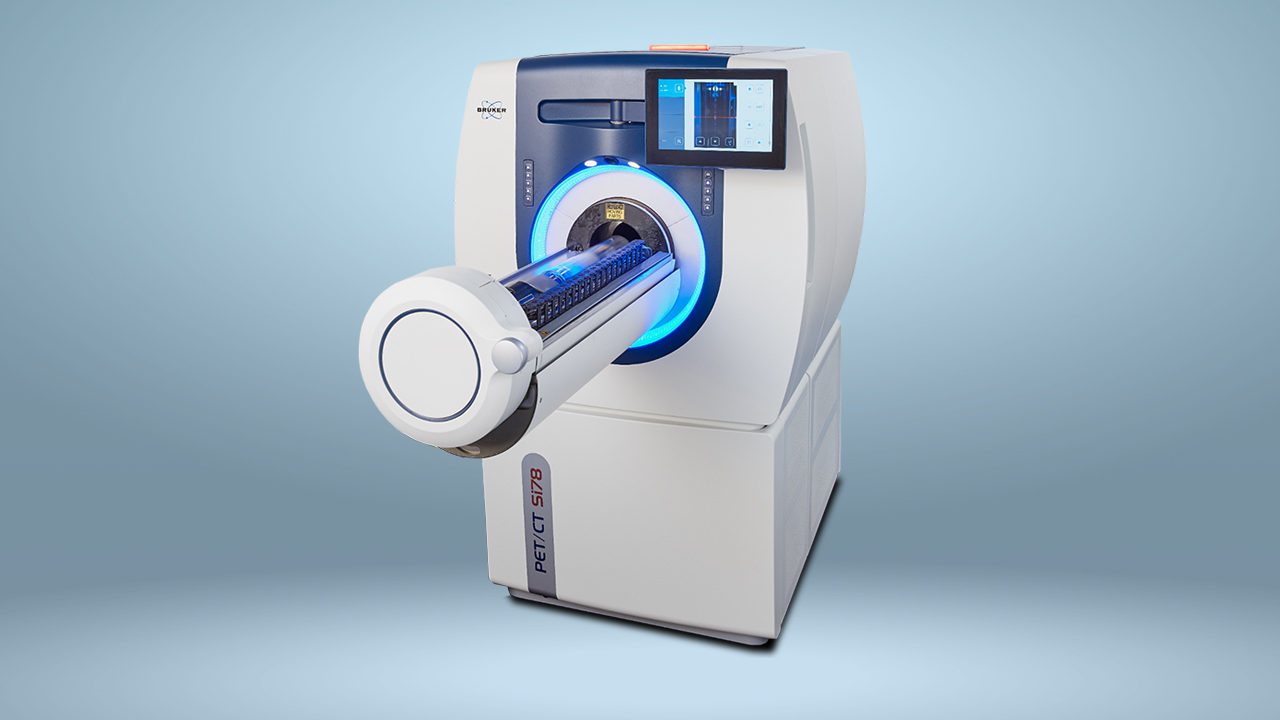
Provide highly sensitive molecular and metabolic imaging of the tumor microenvironment and evaluation of radio-diagnostics and theranostics.
- Micro-CT:
Offers high-resolution anatomical visualization of tumors, bone, and lung structures. Combined with PET/SPECT, CT-supported tissue attenuation can provide highly accurate dosimetry calculations.
Together, these preclinical imaging modalities provide a comprehensive and quantitative understanding of cancer development and treatment response.
Multimodal Preclinical Imaging Solutions for Oncology
Bruker provides a comprehensive portfolio of preclinical imaging solutions for oncology, spanning optical imaging, BLI, FLI, PET, SPECT, MRI, and micro-CT technologies.
These advanced platforms help researchers investigate the mechanisms of tumor progression, evaluate therapeutic efficacy, and accelerate translational cancer research.
Multimodal imaging configurations, including:
- PET/CT
- PET/MR
- SPECT/CT
Coregistration with optical imaging workflows combine molecular, functional, and anatomical information within a single research framework.
By delivering quantitative 3D visualization of radiotracer uptake, tissue morphology, cellular activity, and molecular pathways, Bruker's preclinical imaging solutions empower researchers to advance discoveries that support the development of safer and more effective cancer treatments.