Investigating the Upper Mantle’s Composition and Rheology with Combined Analytical Solutions for Electron Microscopy
Composition and Microstructure Analysis of Peridotite Rocks
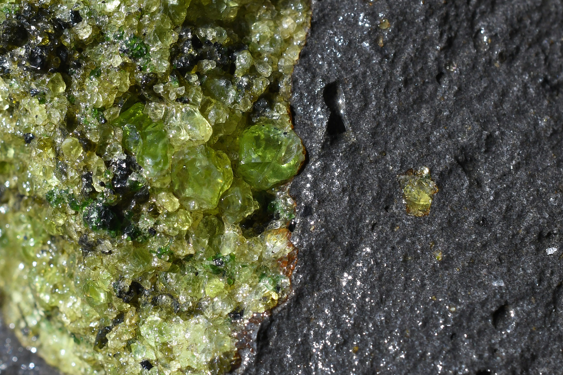
Peridotite rocks are the dominant physical representation of the Earth’s mantle and thus are important to understand geological processes and the Earth’s history. Bruker’s unique ability to combine many analytical techniques to one Scanning Electron Microscope (SEM) allows researchers to comprehensively investigate the composition and microstructure of samples.
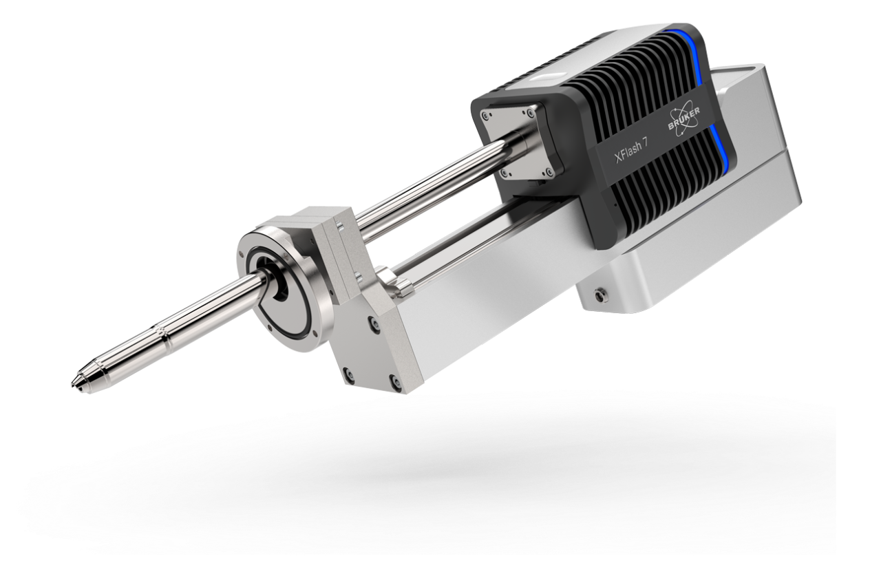
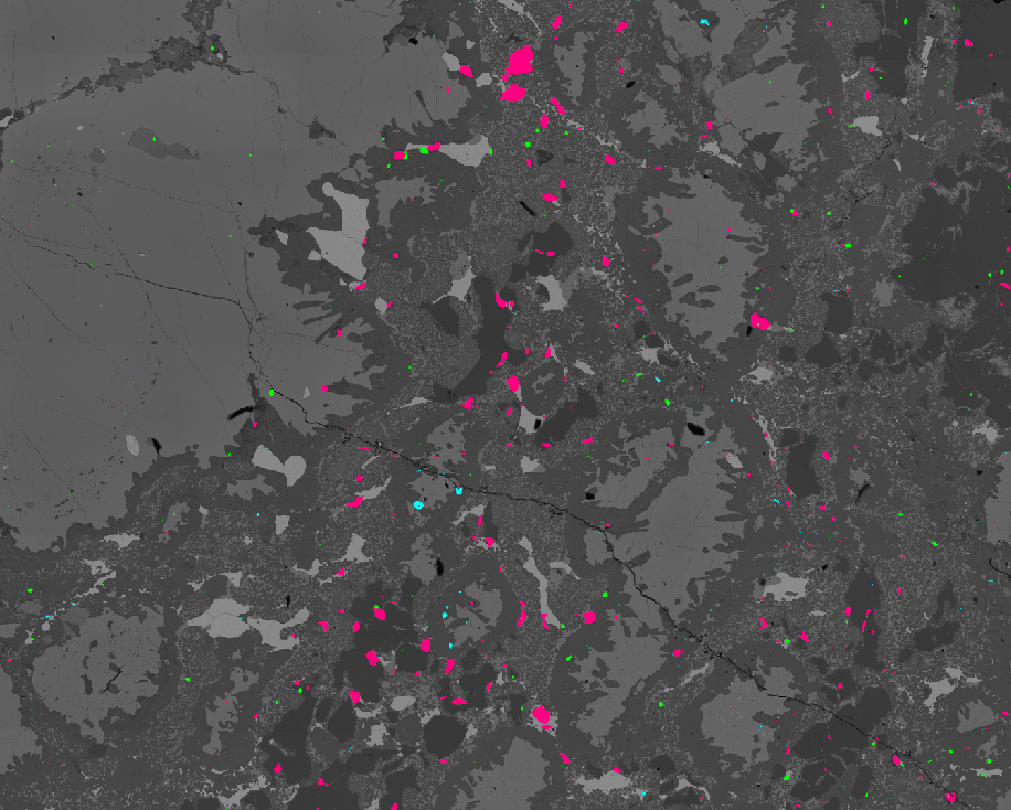
SEM-EDS is a versatile and fast method to spatially resolve the rock composition. The elemental distribution of an entire thin section can be acquired within minutes. This data can be processed with the automated mineralogy software (AMICS) to obtain the mineral identification, distribution, and other relationships.
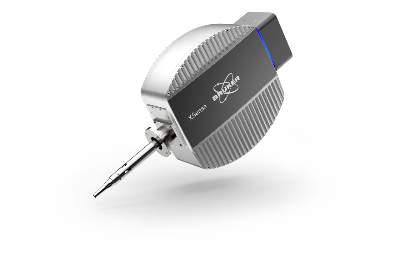
SEM-WDS is the method of choice to identify trace elements with SEM. With its high peak resolution, WDS can identify and quantify trace elements with concentration as low as 100 ppm. In addition, it has the advantage of a considerably higher energy resolution than EDS.
SEM-based micro-XRF analysis provides elemental maps of very large samples. It has low background noise that enables the detection of trace elements as low as 10 ppm and the ability to detect high energy X-ray lines.
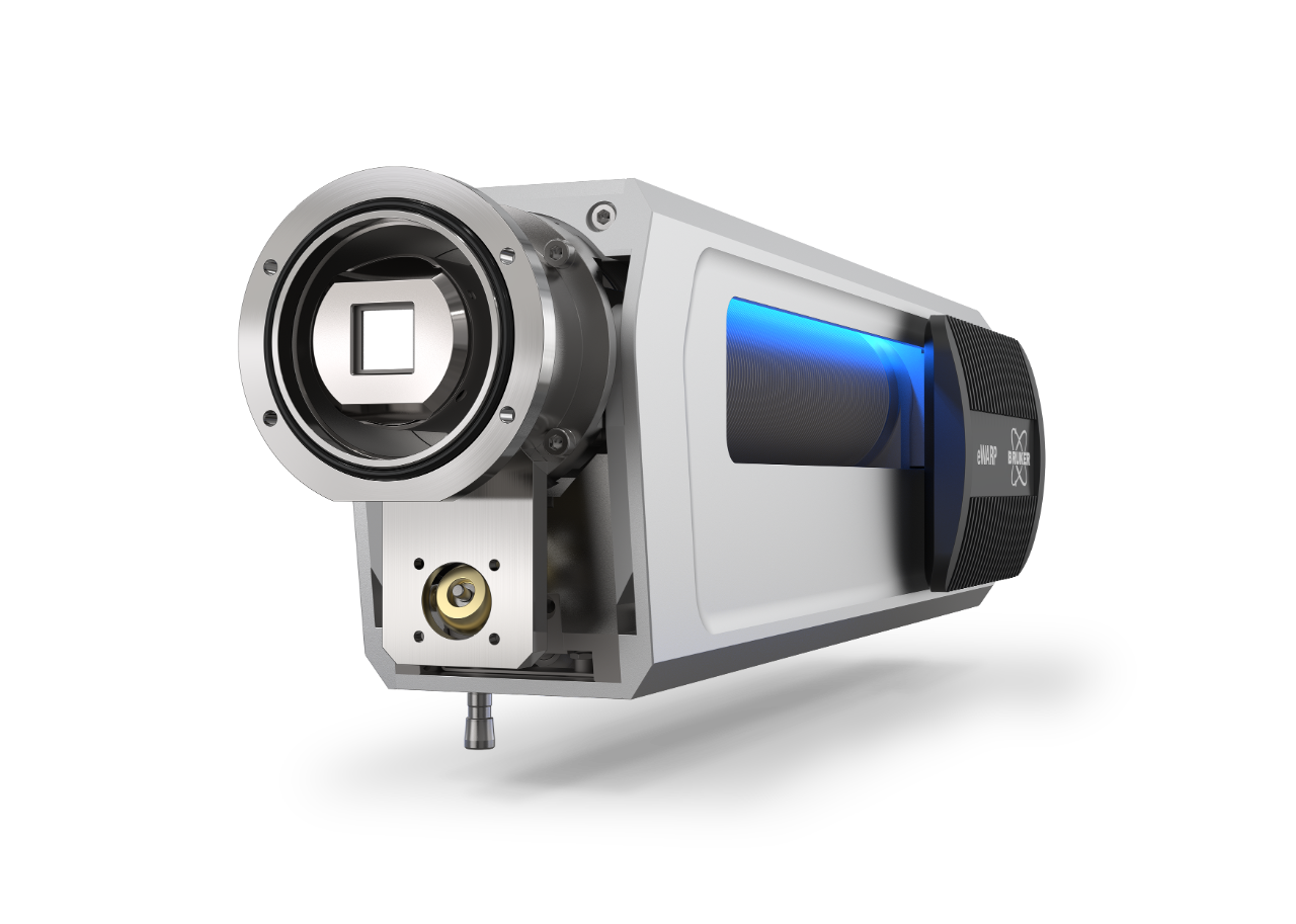
With EBSD, sample microstructure and deformation processes are determined. When combined with EDS, the phases are identified in a semi-automatic way. EBSD is the only technique which correlates microstructure and texture, and thus is an essential technique for completing the characterization of peridotite samples to understand deformation, phase transformation, defects and crystal growth, amongst others.
When combined, these techniques provide the most complete sample analysis possible using a single SEM.


Who Should attend?
- Researchers from academia and mining industries who investigate rocks and their minerals.
- Everyone interested in elemental distribution and microstructure of mineralogical samples.
Speakers

Dr. Katharina Marquardt
Faculty of Engineering, Imperial College, London (UK)

Dr. Michael Abratis
Sr. Application Scientist, Bruker Electron Microscope Analyzers

Dr. Andrew Menzies
Senior Application Scientist - Geology and Mining, Bruker AXS

Dr. Laurie Palasse
Global Application Manager, Bruker Electron Microscope Analyzers
Watch this Webinar On-Demand
Please enter your details below to gain on-demand access to this webinar.