Unlocking 3D Nanostructure in Neurons with Single Molecule Localization Microscopy (SML)
Unlock 3D nanoscale imaging
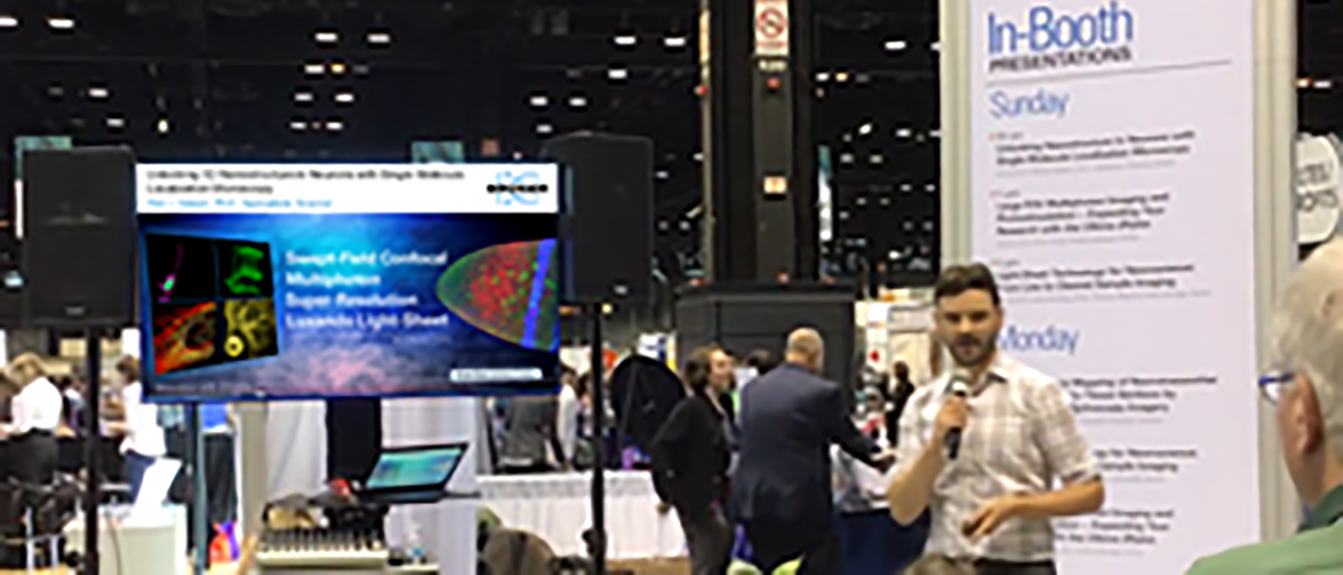
During this webinar, Rob Hobson, Ph.D., a Bruker Applications Scientist, discusses the benefits of SMLM and its applications on the convention hall floor at NEUROSCIENCE 2019. Viewers will gain insights into:
- How single-molecule localization microscopy (SMLM) works
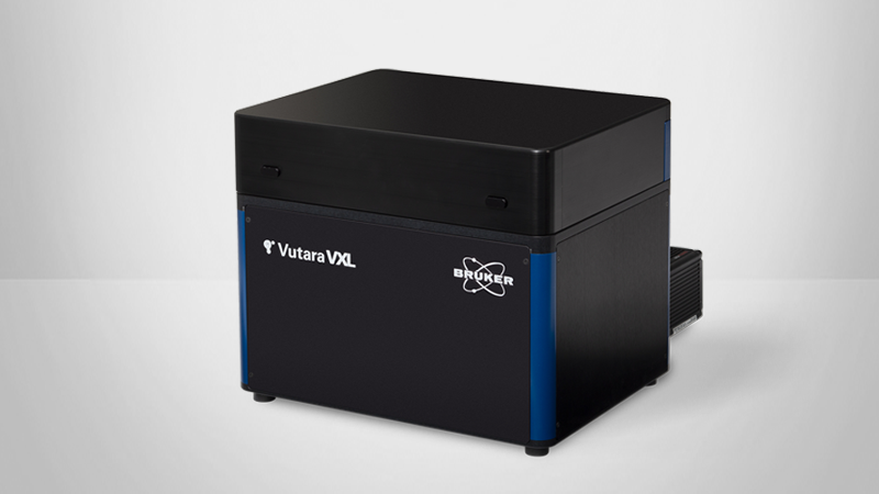
- The Vutara 352 SML super-resolution microscopes and SXR software
- SMLM applications in neuroscience
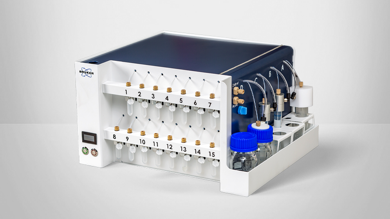
- A new integrated fluidics unit for sequential labeling applications
Webinar Summary
This presentation discusses the principles of single-molecule localization microscopy (SMLM) for overcoming some of the key challenges associated with light microscopy. Our Vutara 352 SML Super-Resolution Microscope and SRX software are uniquely equipped for SMLM applications and advanced neuroscience research in areas such as:
- Infectious diseases
- Cardiology
- Developmental Biology
- Cell Biology
- Neuroscience
Find out more about our other solutions for Super-Resolution Microscopy:
Featured Products and Technology

Speaker

Robert J. Hobson, Ph.D.
Vutara Applications Scientist, Bruker
Dr. Robert Hobson graduated from the University of Salford, UK with a degree in Biology, before completing his PhD in Biology at the University of Toledo, Ohio. He was a research assistant professor at the University of Utah with multiple years’ experience in single-molecule localization microscopy sample preparation and imaging before joining Bruker Nano Surfaces.