
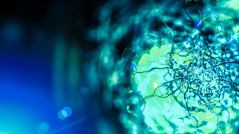
In Vivo Imaging of Microglia as the Brain’s Thermostat for Tuning Neuronal Activity
Discover how top researchers use in-vivo two-photon imaging to explore critical questions in neuroscience research
In this webinar, Dr. Mario Merlini discusses his team's use of in-vivo two-photon imaging methods and other techniques to investigate neuronal excitability. Viewers gain direct insight into:
- The application of in-vivo two-photon imaging for the study of microglial regulation of neuronal activity;
- The two-photon imaging methods used and the techniques that complement them; and
- What this study reveals about the role of microglia dynamics in neuronal hyperexcitability.
Dr. Merlini also answers audience questions about his research including the essential considerations behind his team's experimental approach.
Webinar Summary
Dr. Merlini's research is a great example of the advantages of using two-photon microscopy and techniques like neurotransmitter uncaging, whisker stimulation, and EEG recording for advanced neuroscience research. Watch the on-demand recording or download the webinar recap to find out more.
Presenter's Abstract
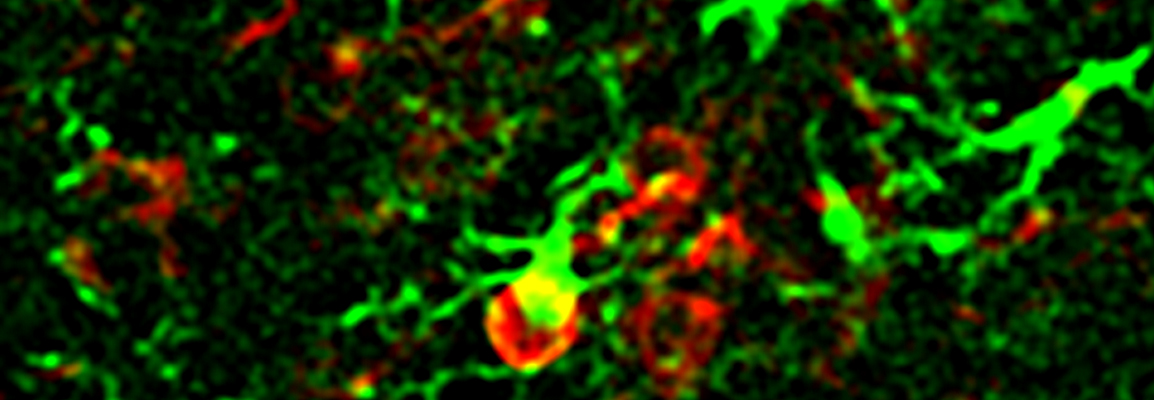
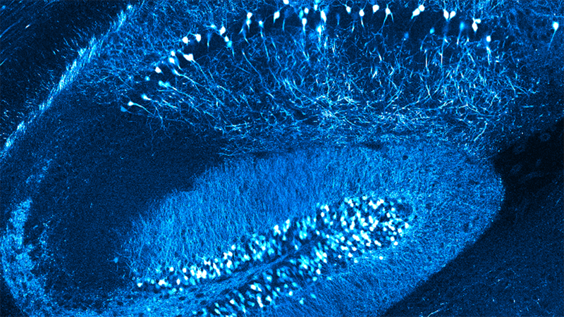
Prevention of neuronal hyperexcitability is essential for healthy brain function, as is illustrated by the aberrant, hyperexcited neuronal activity that contributes to cognitive dysfunction in neurodegenerative diseases and several psychiatric disorders. In this webinar, I will describe recent data acquired with awake in vivo two-photon imaging, in vivo EEG recordings, two-photon-induced glutamate uncaging, and 3D reconstructions of scanning block-face electron microscopy in a newly developed mouse model of chronically impaired microglia motilities.
These mice have hypersynchronized neuronal activity that induces a severe seizure phenotype. The findings reveal a unique role for microglia dynamics in maintaining evoked neuronal activity within a physiological range to prevent brain hyperexcitability, identifying microglia as thermostat-like cells in the regulation of brain activity. I will further present the development of the automated whisker stimulus tool and its integration in a synchronized in vivo two-photon time-lapse image acquisition workflow, as was used in this study for the simultaneous imaging of microglia motilities and evoked intraneuronal Ca2+ transients in awake, head-fixed mice.
Featured Products & Technology
Guest Speaker

Mario Merlini, Ph.D.
Junior Team Leader, Blood & Brain @ Caen Normandie Institute, INSERM U1237, GIP Cyceron, Caen, FranceDr. Mario Merlini is a Junior Team Leader at the Blood & Brain @ Caen Normandie Institute, integrated within the French National Institute of Health and Medical Research (INSERM) and hosted by the biomedical imaging platform Cyceron in Caen, France. He received his Ph.D. degree in neuroscience from the University of Zurich, Switzerland, in the lab of Dr. Roger M. Nitsch studying cerebrovascular and astrocytic pathologies contributing to cognitive impairment in Alzheimer’s disease.
Dr. Merlini was awarded a Swiss National Science Foundation Postdoctoral Fellowship to pursue his postdoctoral research at the Gladstone Institute of Neurological Disease, San Francisco, USA with Dr. Katerina Akassoglou. There, he used in vivo two-photon imaging and 3D imaging in cleared brains to discover a mechanistic link between compromised blood-brain barrier integrity, microglia activation, and dendritic spine loss in Alzheimer’s disease, as well as a fundamental, physiological role for microglia dynamics in tuning neuronal activity. He also developed sensory stimulation and EEG recording methods for application in in-vivo two-photon brain imaging in awake mice. As an independent PI, Dr. Merlini applies his expertise in functional and structural in vivo imaging of glial dynamics, cerebral blood flow, and neuronal activity in experimental animal models and human brains to investigate neurovascular pathways underlying psychiatric disorders and, more fundamentally, how neurovascular interactions shape cognition.