Cellular Mapping of an Entire Organism Using Whole-Body Tissue Clearing and Light-Sheet Microscopy
Explore innovative solutions for whole-body scale fluorescence microscopy
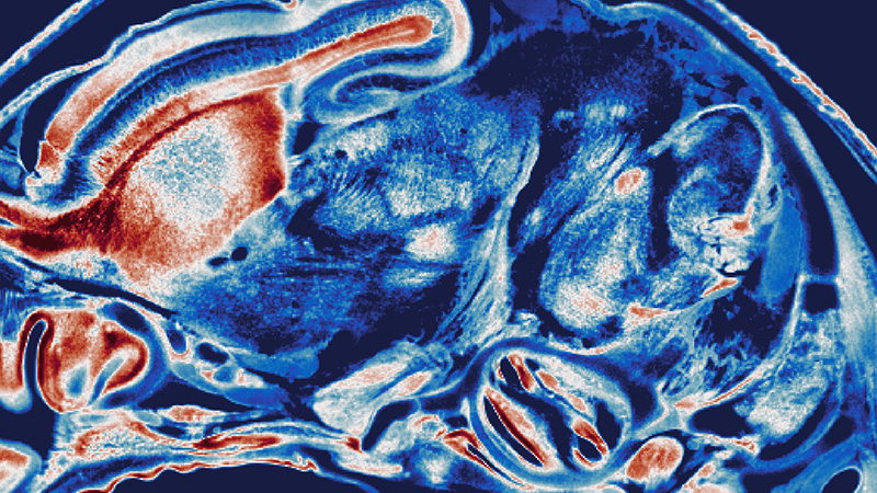
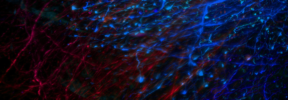
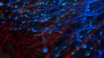
During this webinar, guest speaker Chenchen Pan, Ph.D., University Hospital Heidelberg, discusses his latest research and findings using rodent whole-body scale imaging and deep-learning-based image analysis. Learn how state-of-the-art methods overcome many of the struggles typically associated with organ segmentation for whole-body imaging and quantitative analysis in biomedical research.
Explore Innovative Solutions for Whole-Body Scale Fluorescence Microscopy
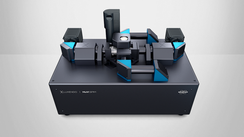
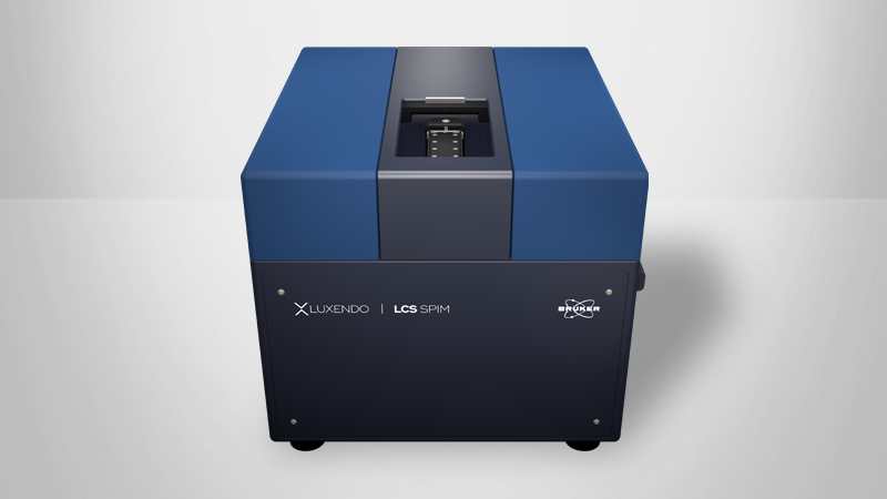
Join us for a one-hour webinar with guest speaker Dr. Chenchen Pan, Department of Neurology, University Hospital Heidelberg. During the live event, Dr. Pan discusses his latest research and findings using rodent whole-body tissue clearing methods combined with light-sheet fluorescence microscopy. He also presents how deep-learning-based image analysis can resolve the cellular-level neuronal connectivity map to achieve cellular mapping of cancer metastasis and therapeutic antibody distribution at a whole-body scale. We also showcase Bruker's light‑sheet microscopes, which provide innovative solutions in sample mounting, sample size, and optics for best-in-class 3D imaging of cleared samples.
Find out more about the technology featured in this webinar or our other fluorescence microscopy solutions: