BioAFM Applications in Cosmetics and Healthcare
Gain deeper insights, streamline development, and strengthen product confidence.
Developing cosmetic and healthcare products requires balancing innovation with practical concerns: enhancing the effectiveness of the product while minimizing adverse side effects and ensuring compliance with healthcare standards. BioAFMs deliver key insights essential to this process, providing nanoscale detail that cannot be resolved by other optical methods, bulk analyses, or classical histology.
Atomic force microscopy measurements for industrial life science applications (BioAFM):
- Provide comprehensive insights into the nanomechanical behavior of biological samples at the cellular and molecular level.
- Reveal key relationships between structure, mechanics, and performance.
- Enable reproducible analysis of live cells, biomaterials, and advanced drug delivery systems.
- Support evidence‑based decisions throughout the development process.
TABLE OF CONTENTS:
LEARN MORE ABOUT THESE APPLICATIONS:
The Technology Behind Better Product Insights
A BioAFM is an atomic force microscope (AFM) adapted, in particular, for the study of biological samples and soft matter. These include fragile and challenging samples such as single molecules, nucleic acids, living cells, and tissues — all under near‑physiological conditions and without staining, labeling, or damaging their structures.
These capabilities help researchers and developers:
- Study the effect of active ingredients on biological matter (e.g., skin, hair, and dental or ocular materials).
- Measure and monitor batch‑to‑batch consistency and product efficacy, safety, and stability.
- Develop and refine advanced biomaterials and delivery systems (e.g., nanoparticles and nanocarriers).
Expand the accordion sections below to learn how:

AFM is a 3D imaging technique that delivers nanometer‑scale resolution, enabling the detection and analysis of surface features and structures that conventional optical methods cannot resolve. These instruments can be used to produce high‑resolution 3D surface profiles, characterize the nanoscale properties of soft materials, living cells, tissues, and cosmetic or healthcare products, and visualize molecular interactions under ambient or near-physiological conditions.
For those working in the cosmetics and healthcare industries, this level of detail helps analyze products and materials, such as creams, serums, contact lenses, oral‑care materials, and hair‑care formulations by revealing subtle structural features that influence product performance, comfort, and tolerance.
BioAFMs enable precise quantification of nanomechanical properties, e.g. stiffness, adhesion, friction, viscoelastic properties, and other parameters that link nanoscale structure to product performance. These measurements can be applied directly to cosmetics and healthcare materials to quantify, for example, friction forces along hair fibers, elasticity in living tissues, collagen stiffness in skin, and the mechanical properties of dentin, enamel, or contact‑lens materials under near‑physiological conditions.
These high‑resolution mechanical datasets support evidence‑based R&D, helping teams optimize formulations, validate performance claims, and ensure product safety and consistency across development and production.
BioAFMs enable label‑free, non‑destructive analysis, with no staining or chemical modification required. Users can perform non-invasive measurements on soft and fragile samples, living cells and tissues, and cosmetic formulations. BioAFMs can measure very small sample quantities, making this technology well suited for the investigation of costly substances, small molecules, and active ingredients used in advanced formulations.
This minimal sample preparation requirement preserves sample authenticity, accelerates testing, and supports both exploratory R&D and routine QC workflows in cosmetics and healthcare development.
BioAFMs can operate under near‑physiological conditions, controlled temperature and humidity, and in liquids, making it possible to study samples in states that reflect real‑world use-cases.
Environmental control enables accurate characterization of hydration effects, lipid organization, surface changes, and biomechanical response, supporting realistic performance testing and more informed formulation decisions across cosmetics and healthcare development.
BioAFM systems incorporate automated workflows and advanced imaging modes such as SmartMapping, enabling large‑area, high‑resolution imaging with nanoscale precision. These automation tools streamline setup, reduce operator‑to‑operator variability, and support high‑throughput analysis of tissues, hair fibers, powders, and other industrial samples.
BioAFM platforms also offer AI‑supported features that further simplify imaging and analysis in industrial environments. Together, these automation capabilities improve reproducibility and throughput, making BioAFM suitable for R&D and quality‑control workflows in cosmetics and healthcare development.
Bruker BioAFMs can be easily combined with advanced optical techniques, e.g., epifluorescence, confocal, STED microscopy, and TIRF, for correlated measurements that enable a more comprehensive sample analysis.
Delivering Insights Across the Cosmetics and Healthcare Production Pipeline
The application areas below show how BioAFMs support R&D, QC, and innovation across the cosmetics and healthcare pipeline. To go straight to specific examples, you can jump to our use‑cases in skincare and dermatology, haircare, dental care, and contact‑lens research.


Product Development
Challenge:
Both industrial R&D teams and academic researchers often work with limited quantities of costly ingredients that require highly sensitive, non-destructive analysis techniques.
Many conventional analytical tools rely on bulk-averaged or purely qualitative measurements, making it difficult to resolve nanoscale properties or interactions and to correlate them reliably with actual product performance.
Solution:
BioAFMs can measure very small sample quantities, making it ideal for the analysis of costly substances, small molecules, and active ingredients used in advanced formulations, providing direct insights into how these interact at the cellular and molecular levels.
Furthermore, high‑speed imaging modes enable users to study dynamic mechanisms, for example, how creams, serums, or active ingredients affect proteins, biomolecules, or cells.
Benefits:
- Accelerate product optimization by revealing nanoscale effects of ingredients on structure, mechanics, and interactions.
- Optimized upstream testing by ruling out weak candidates earlier in the development pipeline.
- Obtain more accurate, true‑to‑life information by preserving sample integrity and enabling measurement under near‑physiological or realistic‑use conditions.
Quality Control & Consistency
Challenge
Many QC methods rely on bulk or destructive tests that cannot reveal variantions in nanoscale properties, surface structure, or biomechnaical characteristics. Rising regulatory demands and consumer expectations are increasing pressure on manufacturers to deliver quantitative, traceable data across raw materials, intermediates, and finished goods.
Solution:
BioAFMs provide high‑resolution, nanoscale surface and mechanical information that helps QC teams detect early indicators of variability before they escalate. The system produces detailed maps of particle morphology, coating uniformity, and viscoelastic response, offering visibility into changes that conventional methods may overlook.
Automated workflows and AI‑supported analysis enable repeatable, operator‑independent measurements and make nanoscale metrology practical for real‑time quality assessment.
Benefits:
- Detect subtle nanoscale changes in structure, coating uniformity, or mechanical response before they affect product performance.
- Support quality compliance with quantitative, traceable data.
- Improve customer loyalty with consistent product quality and evidence-based claims.
Innovation & Personalization
Challenge:
A new generation of cosmetic and healthcare products relies on tailored performance. As product lines become more targeted, R&D teams increasingly depend on nanoscale evidence to differentiate performance, validate claims, and design customized solutions.
Solution:
BioAFMs provide high‑resolution imaging and quantitative nanomechanical measurements under near‑physiological or controlled environmental conditions. It supports label‑free analysis of soft and living samples, integrates with optical or spectroscopic techniques, and incorporates automated workflows such as SmartMapping, for large‑area, nanoscale‑precision measurements. These capabilities allow teams to compare formulation variants or new material concepts with quantitative detail that conventional tools cannot provide.
Benefits:
- Support development of personalized or high‑specificity formulations with nanoscale structural and biomechanical evidence.
- Quantitative analysis of product performance based on mechanical properties, such as texture, adhesion, stiffness, and surface behavior.
- Provide the data needed to substantiate performance claims for innovative or tailored cosmetic and healthcare products.
How BioAFMs Are Being Used
From skincare and dermatology to haircare, dental treatments and contact lenses, BioAFMs can be used in the development of a wide range of cosmetic and healthcare materials. The summaries below highlight how atomic force microscopy supports product development, quality assessment, and performance optimization in these areas, offering label‑free, high‑resolution insights under realistic conditions.
For a deeper look at these use-cases, you can download the accompanying Application Note: BioAFM in the Cosmetic and Healthcare Industries.
Skincare & Dermatology
Atomic force microscopy measurements provide a nanoscale view of skin structure and mechanics that goes beyond traditional histology. By combining high‑resolution topography measurements with quantitative nanomechanical mapping, researchers can evaluate collagen structure, tissue stiffness, and the effects of care products on skin cells in environments that closely resembles real-life biological conditions.
These capabilities help developers and clinicians:
- Distinguish subtle structural and mechanical alterations.
- Evaluate product performance and the efficacy of treatments.
- Generate evidence-based claims in personalized skincare and therapeutic formulations.

In one study, researchers used BioAFM technology to examine dermal tissue sections from healthy donors and patients with hEDS and scleroderma.
(A) Correlated AFM and PS-stained images, identifying intact and compromised regions within skin samples obtained from control and hEDS-Scleroderma patients. (B) Collagen fibrils' Young's moduli distributions for healthy and disrupted areas. The red dotted line presents the healthy collagen distribution in the control group. Data courtesy Laurent Bozec University of Toronto, Canada.

In a representative study, AFM was used to scan individual hair fibers before and after applying a conditioning product.
FIGURE 3. AFM analysis of a hair fiber before and after application of a conditioning hair care product. AFM topography images (left) of a hair fiber and corresponding friction measurements (right) along the hair fiber (indicated by the red line). The diagrams show the leveled height profile in blue and the corresponding friction forces: purple (trace) and green (retrace). A: Untreated hair fiber. Topography image (A1) and corresponding friction measurement (A2). B: Hair fiber after treatment with conditioner. Topography image (B1) and corresponding friction measurement (B2).
Hair Care
The cuticle surface governs friction, adhesion, and wear — properties that directly influence smoothness, manageability, and overall feel. Atomic force microscopy can be used to measure tribological behavior under specific conditions and in controlled environments, offering insights beyond visual and macroscopic assessment.
These datasets support:
- Performance comparison across formulations.
- Validation of “smoothness” or “reduced friction” claims.
- R&D studies exploring the effectivity of cuticle protection, hydration, and repair.
Dental Care
For tooth care products, the challenge is balancing cleaning or whitening effectiveness with enamel and dentin preservation. Atomic force microscopy allows developers to evaluate abrasives, bleaching agents, desensitizing ingredients, and protective coatings by visualizing and quantifying nanoscale changes in tooth structure and mechanics.
These measurements help teams:
- Compare erosive or protective effects across formulations.
- Visualize and quantify dentin tubule sealing in desensitizing products.
- Support claims such as “protects enamel,” or “reduces sensitivity” with evidence.

In the experiment featured in our application note "BioAFM in the Cosmetic and Healthcare Industries," AFM topography measurements revealed how dentin surface morphology changed after brushing and exposure to a bleaching gel — capturing structural changes to the integrity of the enamel.
FIGURE 4. AFM analysis of animal tooth cross-section. The sample was prepared by sanding, polishing, and etching with 0.5M EDTA for 2 minutes. A: AFM topography image of the dentin surface showing dentin tubules (Z-range: 618 nm). B: Zoomed in AFM topography image of dentin surface after brushing with toothpaste for 30 min and subsequent cleaning with water (Z-range: 150 nm). C: AFM topography image of dentin surface after application of bleaching gel, revealing structural changes (Z-range: 150 nm).

In a representative study, AFM was applied to daily disposable silicone hydrogel lenses. Using SmartMapping, millimeter‑scale curvature maps were generated with nanoscale precision, revealing variations across the lens that relate to comfort and fit.
FIGURE 1. AFM measurements on daily contact lenses (single-use): A: Large scale topography map (Z-range: 90 μm). B: Corresponding Young’s modulus map (Z range: 326-506 kPa; average modulus: 405 kPa), acquired using SmartMapping mode. C, D: High-resolution AFM topography images of the lens surface. E: Frequency-dependent analysis of viscoelastic properties showing Storage Modulus (E’), Loss Modulus (E”), and Loss Tangent (ratio of E”/E’).
Contact Lenses
BioAFM enables nanoscale evaluation of lens materials, coatings, and surface‑treatment technologies in fluid or under near‑physiological conditions. By combining high‑resolution imaging with quantitative nanomechanical mapping, teams can:
- Analyze surface topography, stiffness, and viscoelastic characteristics relevant to comfort and wearability.
- Assist the development of specialized materials and surface coatings that reduce irriation and the risk of infection.
- Support quality assurance during the manufacturing process.
These measurements help developers to improve the comfort, durability, and overall ocular compatibility of contact lenses by providing quantitative data that traditional optical methods often cannot deliver.
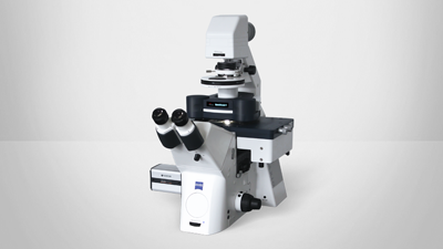
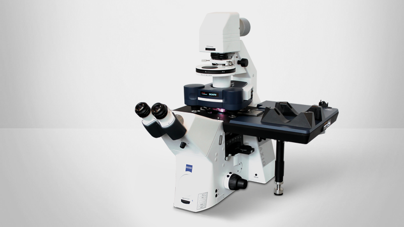
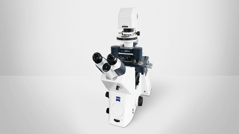
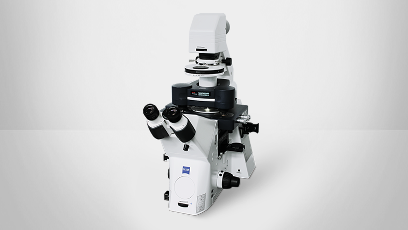
BioAFMs for the Cosmetics and Healthcare Industries
All Bruker BioAFM systems can support cosmetics and healthcare applications. Based on the workflows, automation needs, and nanoscale characterization strategies discussed by our application scientists, we recommend that interested users start by exploring the systems highlighted below, which align most closely with the studies and industrial use‑cases described above.