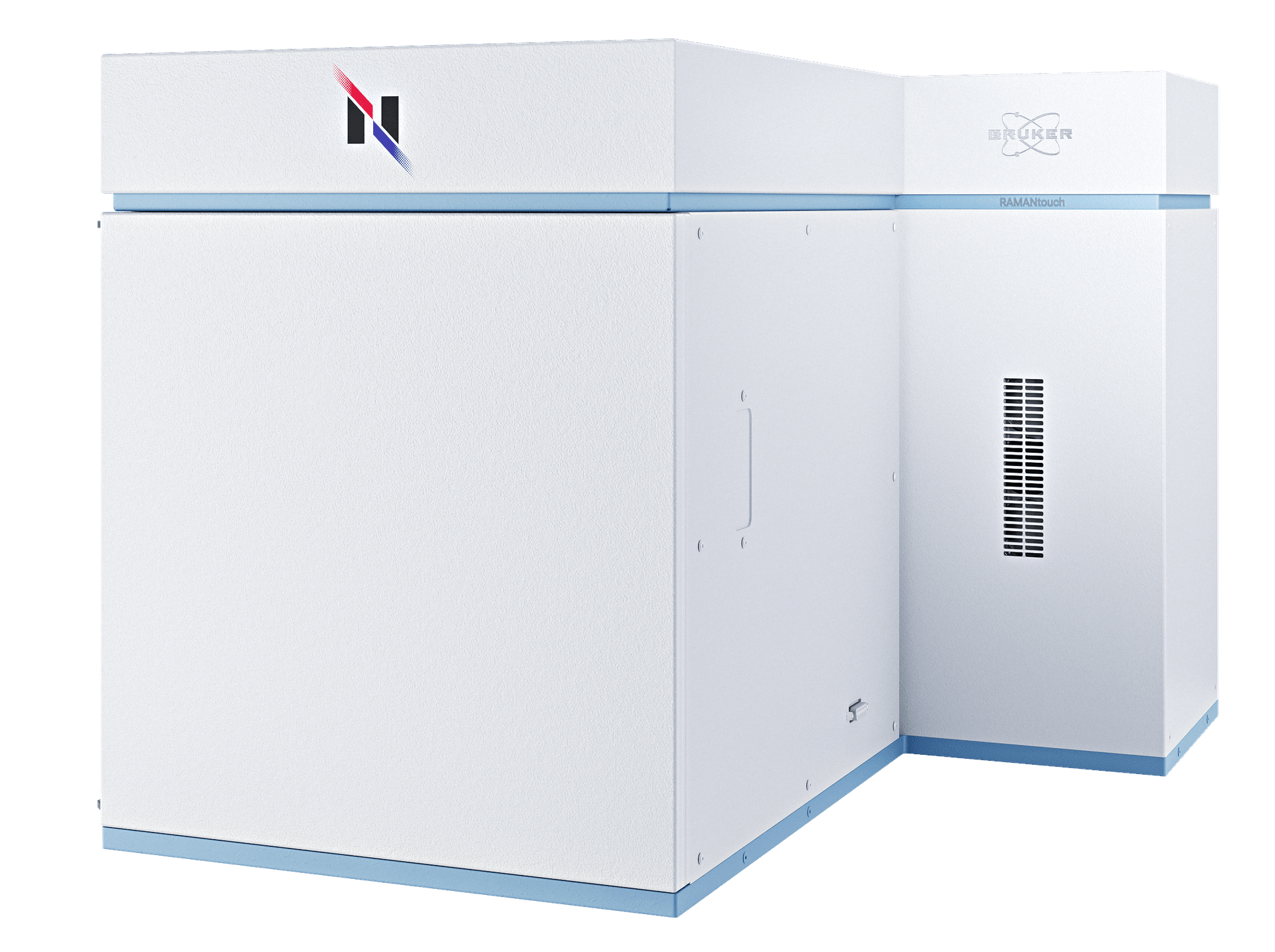
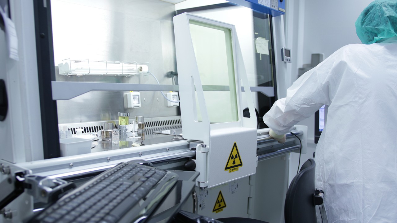
RAMANtouch
Et Voici
Le Raman imagerie le plus rapide
Un microscope d'imagerie confocale Raman avec une vitesse et une précision inégalées
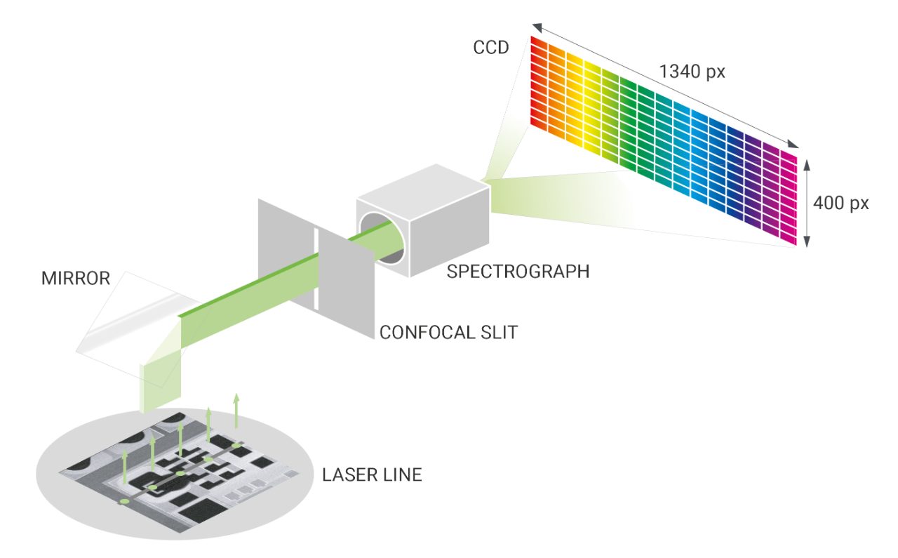
La technologie de Nanophoton utilise un éclairage laser en forme de ligne et un CCD bidimensionnel pour capturer 400 spectres en une seule exposition. Grâce à la numérisation laser via des scanners galvanométriques, RAMANtouch permet d'obtenir une imagerie Raman rapide et précise de centaines de milliers de pixels en quelques minutes - aucun EMCCD n'est requis.
La véritable imagerie Raman est enfin là
Le RAMANtouch est une approche totalement nouvelle par rapport aux microscopes Raman traditionnels - et c'est pourquoi il réalise l'impossible. Une vitesse d'imagerie maximale sans aucune perte de qualité spectrale ou de résolution spatiale.
Le RAMANtouch offre :
- Imagerie Raman ultra rapide
- Haute résolution spectrale et spatiale
- Système entièrement automatisé
- Analyse puissante pour les images Raman 2D/3D
- Calibrage et alignement automatiques

stay tuned for more !
Explication du balayage laser par galvanomètre
L'utilisation d'un miroir galvanométrique est une méthode de balayage innovante dans laquelle un faisceau laser peut balayer librement le point situé sous l'objectif - sans avoir à déplacer la platine de l'échantillon ! En termes de précision et de rapidité, il surpasse de loin le "balayage sur table" conventionnel.
De plus, le faisceau laser est incident et perpendiculairement au plan d'observation, que vous soyez au centre du champ de vision ou sur le bord. Cela permet un mode de mesure spécials:
Balayage de lignes laser :
Numérisez la surface de votre échantillon avec une résolution spectrale et spatiale élevée à la vitesse la plus élevée et avec une précision maximale.
Numérisation de points :
Créez des images d'aperçu rapides d'une zone de mesure en mesurant uniquement les parties qui présentent réellement un contraste dans leurs spectres Raman..
Mesure AreaFlash :
En faisant clignoter la ligne laser Raman sur le champ de vision, un spectre moyen de la zone complète est créé. Cela permet des mesures ultra-rapides sur de grandes surfaces.
Performances Raman maximales dans n'importe quel mode de mesure
Mode ligne Laser
- Imagerie Raman ultra-rapide par illumination linéaire
- Ligne laser homogène et sans déformation sur tout le champ de vision
- Aucun emCCD nécessaire
Mode point Laser
- Mise au point parfaite même au bord du champ de vision
- Des centaines de fois plus rapide qu'une platine motorisée
- Précision de positionnement jusqu'à 10 nm
Capacités exceptionnelles d'imagerie Raman 3D
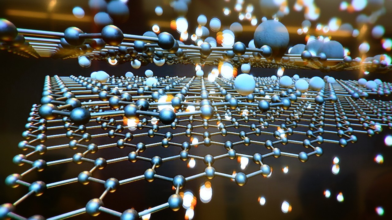
L'optique confocale permet une analyse Raman non destructive à l'intérieur d'un échantillon. De cette façon, des images Raman 3D d'échantillons transparents peuvent être créées. En utilisant l'éclairage linéaire et la platine haute précision du RAMANtouch, des images Raman 3D sont créées avec une vitesse et une qualité inégalées pour fournir un aperçu de la structure interne et de la distribution des composants d'un échantillon.
Exemples en Vidéo :
- Détection Raman à l'intérieur de la fibre transparente
- Échantillon : fibre bicomposante PE (gaîne) et PET (coeur)
- Durée d'acquisition : 20 ~ 30 min
- Yellow dots: TiO2
Résolution maximale de l'imagerie Raman
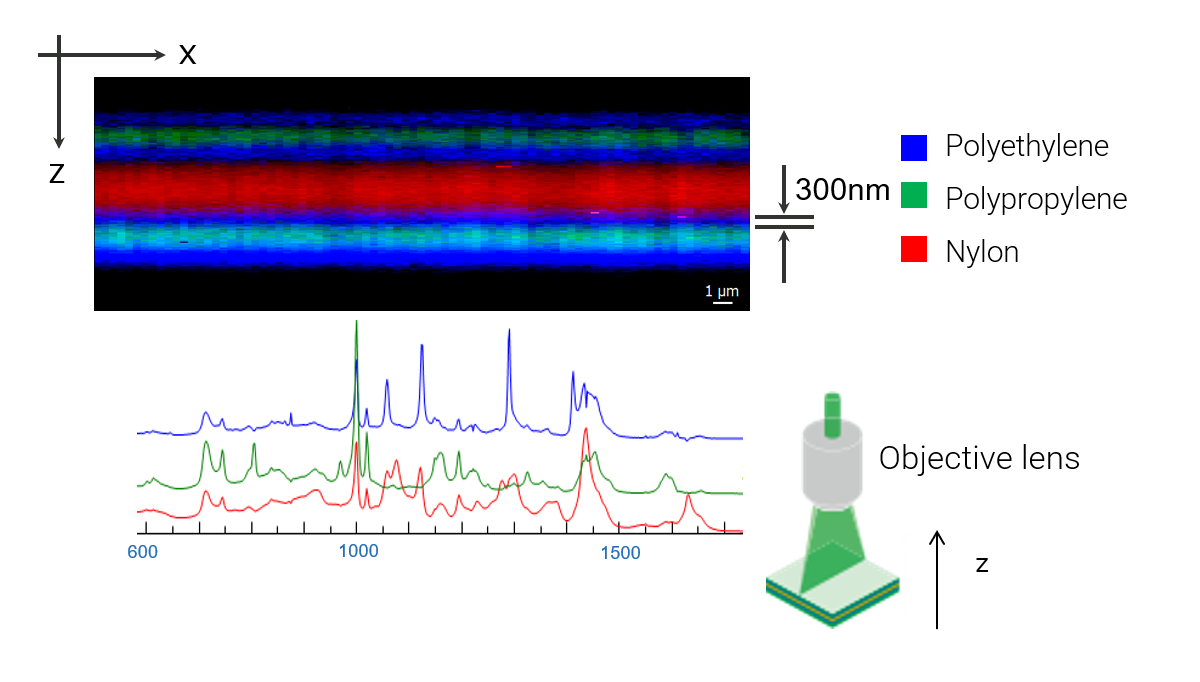
L'imagerie Raman à profil de profondeur dans le plan XZ permet une analyse non destructive, comme l'examen de films multicouches. L'utilisation d'un objectif à immersion dans l'huile améliore considérablement la résolution spatiale, permettant la détection de couches ultra-minces aussi fines que 250 nm.
Accéder au domaine submicrométrique
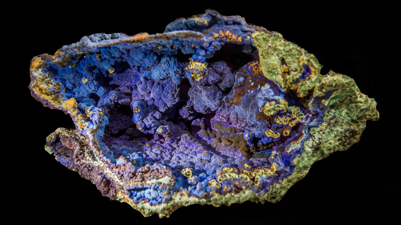
La microscopie Raman offre des informations essentielles sur l'identification des composés organiques et inorganiques, ainsi que sur le polymorphisme cristallin. Contrairement à la microscopie IR, la microscopie Raman présente des avantages tels qu'une résolution spatiale inférieure au micromètre et la capacité d'analyser des échantillons dans des environnements aqueux.


Spécifications RAMANtouch
| Spatial Resolution | 350 nm in X, 500 nm in Y; 1 µm in Z | |
| Objective Lenses | 5x, 10x, 20x, 50x, 100x | |
| Spectral Resolution | <0.9 cm-1 (depends on grating, up to 3 gratings available) | |
| Stage Details | 30 * 30 * 35 mm XYZ-motorized stage | |
| Calibration | Auto-calibration based on standard lamp and sample | |
| Alignment | Auto-alignment of optical path | |
| Laser Safety | Laser safety class I door with interlock |

When I first encountered a Bruker’s Nanophoton Raman microscope, which was installed for the first time at Analytical center in Chungbuk National University, I was very surprised by the various performances that are different from the previous Raman microscopy. In particular, I was very impressed the function of large-area mapping, which is a disadvantage of most of current Raman microscopy, was solved with the idea of line-illumination.
In our laboratory, Raman spectroscopy was utilized to quantify amino acids in renal cell cultures from an animal model of induced renal dysfunction and to study the distribution of drugs on the cell membrane surface. In addition, the content of illicitly distributed APIs-like compounds was determined by Raman imaging and a semi-quantification method was established.
In addition, the bio-samples that we are mainly researching in our laboratory are very difficult to find the optimum experimental conditions due to the control of the laser power, but the RAMANtouch model can not only adjust a very small power level, but also use the preview function to find the optimal conditions faster than any other Raman product. By using such a powerful Raman microscopy in our laboratory, we have published several number of related papers.
Professor Dr. Yong-Moon Lee, School of Pharmacy, Chungbuk National University, Korea.

The RAMANtouch microscope has made us rethink the limits of application of Raman imaging. The multiplexed acquisition approach brings a significant increase in speed, making it feasible to collect large Raman images instead of few measurement points. The system automation enables our users to utilize measurement time efficiently in overnight runs. This capability adds analytical depth to Raman characterization and facilitates a better understanding of complex research questions and enables big data exploration with machine learning tools.
At Synchrotron Soleil, we facilitate scientific progress by providing access to a high level research infrastructure. Beyond the beamline instrumentation, we also provide additional characterization tools such as offline Infrared or Raman microscopy.
Ferenc Borondis, Beamline Manager & Principal Beamline Scientist at Synchrotron SOLEI, Saint-Aubin, France.

Now, with the pioneering application of the modifed RAMANtouch system to the world’s first 8.5th generation display mass production line, we can achieve groundbreaking real-time defect analysis directly within the production process. By enabling component-level analysis of random defects—responsible for over 90% of yield-loss issues—this technology allows for precise defect identification, helping manufacturers not only trace defect origins and specify affected processes but also prevent these issues in real time.
In the high-stakes realm of display product mass production, advanced inspection technology has become essential for optimizing yield and ensuring top-tier quality control. As these technologies evolve, the need for faster, more precise process feedback has driven demand for real-time monitoring that goes beyond traditional inspection to include advanced measurement and in-depth analysis. Furthermore, In-line customized RAMANtouch’s proactive feedback capabilities offer unprecedented opportunities for continuous process improvement and accident prevention, marking a transformative step forward in display production technology.
Dr. Yong-Woon Lim, Metrology & Inspection Team, Samsung Display, Korea.
Frequently Asked Questions
- Why does RAMANtouch achieve high spatial resolution?
RAMANtouch is designed strictly according to optical principles and assembled by specialists in optical design, which ensures that every unit achieves stable and reproducible performance. Its compact mechanical structure also minimizes the likelihood of misalignment caused by environmental changes. - Do I need to adjust the optics when switching lasers?
No manual adjustments are necessary. When a laser is switched, the required optical adjustments are performed automatically, and the change can be carried out with a single click. - Do I need to adjust the optics when switching diffraction gratings?
No. Just like laser switching, the instrument automatically adjusts the optics after the grating is changed, and the user does not need to perform any manual alignment. - Why are line illumination and laser beam scanning used?
These methods reflect Nanophoton’s origins in laser microscopy. Laser beam scanning eliminates the need to adjust measurement positions by moving a motorized stage and avoids issues such as vibration or sample displacement introduced by stage movement. The proprietary line illumination method enables the acquisition of 400 Raman spectra in the X-direction with a single irradiation, allowing high-speed Raman imaging. - How is illumination unevenness in line illumination addressed?
Although uneven intensity between the center and edges of the line is a common concern in line-illumination systems, RAMANtouch significantly reduces this issue using a proprietary technology covered by U.S. Patent US7561265. - Can high-power laser irradiation damage samples?
RAMANtouch includes an ND filter that provides nearly stepless control of laser power across 256 levels. This allows high power to be used efficiently for samples that tolerate it, while also enabling safe measurement of heat-sensitive samples by reducing the power appropriately. - How large is the field of view?
The field of view depends on the objective lens. With a 20× objective, the observation area is about 400 µm × 600 µm and the maximum imaging range is approximately 400 µm square. With a 100× objective, the observation area is roughly 80 µm × 100 µm and the maximum imaging range is about 80 µm square.


