RAMANtouch
Rethink
Raman Imaging
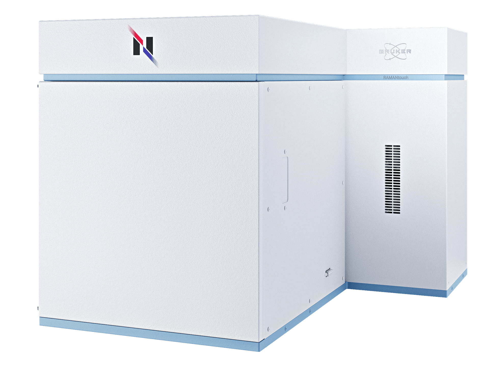
兼具速度和精度的共聚焦拉曼成像显微镜
Nanophoton拉曼显微镜采用线形激光照明和二维CCD技术,一次曝光即可得到400个光谱。通过振镜扫描仪进行激光扫描时,RAMANtouch可在几分钟内,快速、准确地实现数十万像素的拉曼成像——无需电子倍增CCD(EMCCD)。
真正的拉曼成像终于到来
与传统拉曼显微镜相比,RAMANtouch采用了一种全新的分析方法——这也是它化不可能为可能的原因:在实现最高成像速度的同时,不损失任何光谱质量或空间分辨率。
RAMANtouch具有以下优势:
- 超高速拉曼成像
- 较高的光谱和空间分辨率
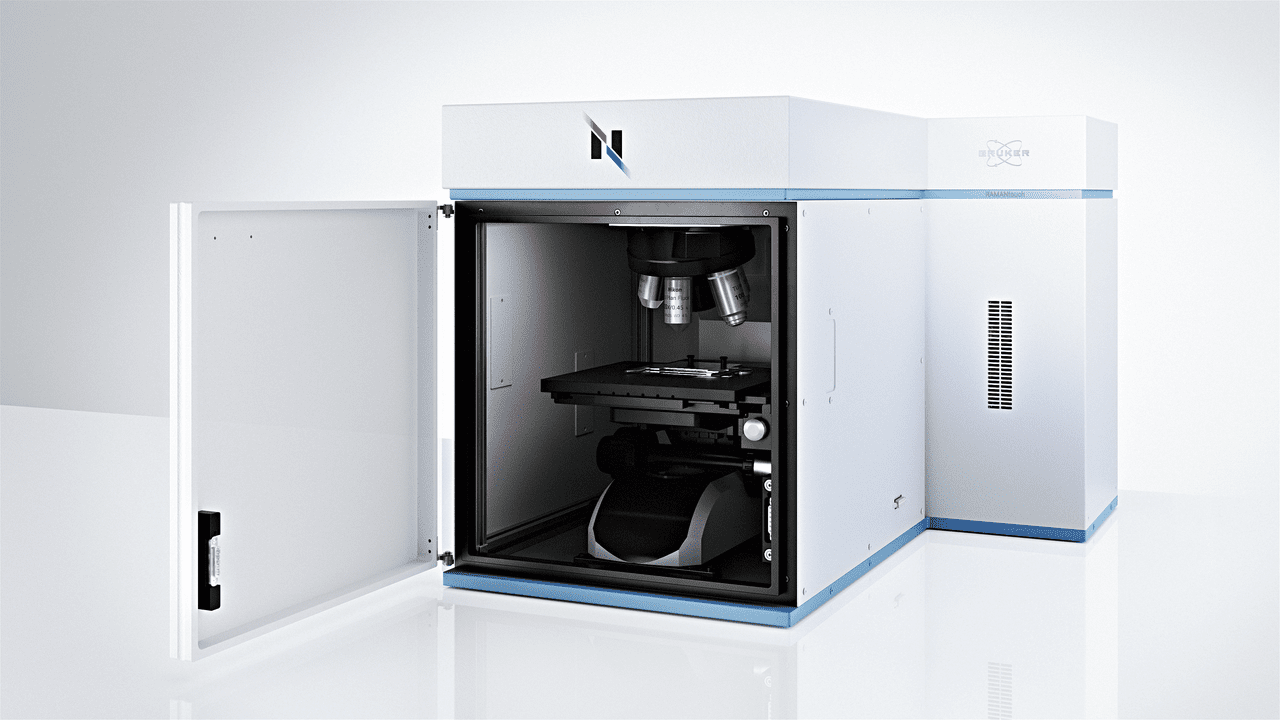
- 完全自动化的硬件
- 强大的2D/3D拉曼图像分析
- 自动校准和自动准直
stay tuned for more !
振镜扫描仪激光扫描
RAMANtouch使用了振镜,这是一种创新的扫描方法,使激光束可以在物镜下自由扫描目标点——无需移动样品台!不论是精度,还是速度,它都远远超过了传统的“移动台面进行扫描”。
此外,无论您位于视野中心,还是在边缘,激光束都垂直于观察平面入射。这一特点可以实现特殊的测量模式:
- 激光线扫描
以最快的速度和最高的精度,对样品表面进行高光谱和空间分辨率扫描。
- 点扫描
通过仅测量拉曼光谱中显示对比度的部分,快速创建测量区域的概览图像。
- AreaFlash测量
通过在视野(FOV)上“闪烁”拉曼激光线,创建整个区域的平均光谱。这一特点使得RAMANtouch可以对大面积区域进行超快速测量。

在任何测量模式下都能最大限度地发挥拉曼性能
激光线模式
- 通过线照明,实现超快速拉曼成像
- 整个视野范围内的激光线均匀且无变形
- 无需电子倍增CCD
激光点模式
- 即使在视野边缘也能实现理想的聚焦
- 比电动样品台快数百倍
- 定位精度高达10纳米
优异的3D拉曼成像能力
共聚焦光学系统可以在样品内部进行非破坏性拉曼分析。通过这种方式,可以对透明样品创建3D拉曼图像。借助线照明和RAMANtouch的高精度平台,您将能以出色的速度和质量,创建3D拉曼图像,从而深入了解样品的内部结构和成分分布。
视频示例:
- 使用拉曼显微镜检测透明纤维内部
- 样品: PE(包层)和 PET(芯)双组分纤维
- 采集时间:20~30分钟
- 黄点:TiO2
峰拉曼成像分辨率
在XZ平面上进行深度剖面拉曼成像,可实现非破坏性分析,例如,检查多层薄膜。利用油浸物镜可显著提高空间分辨率,能检测到厚度仅为250纳米的超薄层。

进入亚微米领域
拉曼显微镜为识别有机和无机化合物以及晶体多态性提供了重要洞见。不同于红外显微镜,拉曼显微镜具有亚微米级的空间分辨率,以及在水相环境中分析样品等优势。


RAMANtouch技术参数
| Spatial Resolution | 350 nm in X, 500 nm in Y; 1 µm in Z | |
| Objective Lenses | 5x, 10x, 20x, 50x, 100x | |
| Spectral Resolution | <0.9 cm-1 (depends on grating, up to 3 gratings available) | |
| Stage Details | 30 * 30 * 35 mm XYZ-motorized stage | |
| Calibration | Auto-calibration based on standard lamp and sample | |
| Alignment | Auto-alignment of optical path | |
| Laser Safety | Laser safety class I door with interlock |

When I first encountered a Bruker’s Nanophoton Raman microscope, which was installed for the first time at Analytical center in Chungbuk National University, I was very surprised by the various performances that are different from the previous Raman microscopy. In particular, I was very impressed the function of large-area mapping, which is a disadvantage of most of current Raman microscopy, was solved with the idea of line-illumination.
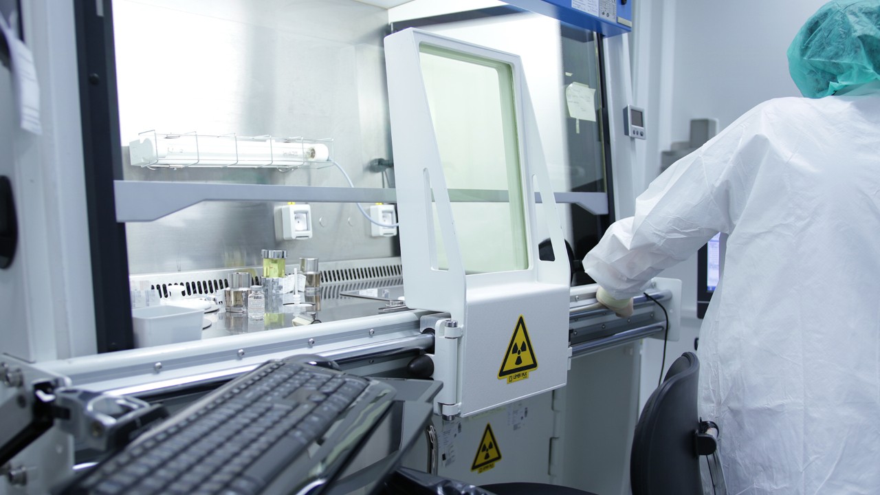
In our laboratory, Raman spectroscopy was utilized to quantify amino acids in renal cell cultures from an animal model of induced renal dysfunction and to study the distribution of drugs on the cell membrane surface. In addition, the content of illicitly distributed APIs-like compounds was determined by Raman imaging and a semi-quantification method was established.
In addition, the bio-samples that we are mainly researching in our laboratory are very difficult to find the optimum experimental conditions due to the control of the laser power, but the RAMANtouch model can not only adjust a very small power level, but also use the preview function to find the optimal conditions faster than any other Raman product. By using such a powerful Raman microscopy in our laboratory, we have published several number of related papers.
Professor Dr. Yong-Moon Lee, School of Pharmacy, Chungbuk National University, Korea.

The RAMANtouch microscope has made us rethink the limits of application of Raman imaging. The multiplexed acquisition approach brings a significant increase in speed, making it feasible to collect large Raman images instead of few measurement points. The system automation enables our users to utilize measurement time efficiently in overnight runs. This capability adds analytical depth to Raman characterization and facilitates a better understanding of complex research questions and enables big data exploration with machine learning tools.
At Synchrotron Soleil, we facilitate scientific progress by providing access to a high level research infrastructure. Beyond the beamline instrumentation, we also provide additional characterization tools such as offline Infrared or Raman microscopy.
Ferenc Borondis, Beamline Manager & Principal Beamline Scientist at Synchrotron SOLEI, Saint-Aubin, France.

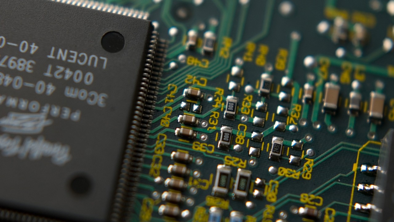
Now, with the pioneering application of the modifed RAMANtouch system to the world’s first 8.5th generation display mass production line, we can achieve groundbreaking real-time defect analysis directly within the production process. By enabling component-level analysis of random defects—responsible for over 90% of yield-loss issues—this technology allows for precise defect identification, helping manufacturers not only trace defect origins and specify affected processes but also prevent these issues in real time.
In the high-stakes realm of display product mass production, advanced inspection technology has become essential for optimizing yield and ensuring top-tier quality control. As these technologies evolve, the need for faster, more precise process feedback has driven demand for real-time monitoring that goes beyond traditional inspection to include advanced measurement and in-depth analysis. Furthermore, In-line customized RAMANtouch’s proactive feedback capabilities offer unprecedented opportunities for continuous process improvement and accident prevention, marking a transformative step forward in display production technology.
Dr. Yong-Woon Lim, Metrology & Inspection Team, Samsung Display, Korea.
Frequently Asked Questions
- Why does RAMANtouch achieve high spatial resolution?
RAMANtouch is designed strictly according to optical principles and assembled by specialists in optical design, which ensures that every unit achieves stable and reproducible performance. Its compact mechanical structure also minimizes the likelihood of misalignment caused by environmental changes. - Do I need to adjust the optics when switching lasers?
No manual adjustments are necessary. When a laser is switched, the required optical adjustments are performed automatically, and the change can be carried out with a single click. - Do I need to adjust the optics when switching diffraction gratings?
No. Just like laser switching, the instrument automatically adjusts the optics after the grating is changed, and the user does not need to perform any manual alignment. - Why are line illumination and laser beam scanning used?
These methods reflect Nanophoton’s origins in laser microscopy. Laser beam scanning eliminates the need to adjust measurement positions by moving a motorized stage and avoids issues such as vibration or sample displacement introduced by stage movement. The proprietary line illumination method enables the acquisition of 400 Raman spectra in the X-direction with a single irradiation, allowing high-speed Raman imaging. - How is illumination unevenness in line illumination addressed?
Although uneven intensity between the center and edges of the line is a common concern in line-illumination systems, RAMANtouch significantly reduces this issue using a proprietary technology covered by U.S. Patent US7561265. - Can high-power laser irradiation damage samples?
RAMANtouch includes an ND filter that provides nearly stepless control of laser power across 256 levels. This allows high power to be used efficiently for samples that tolerate it, while also enabling safe measurement of heat-sensitive samples by reducing the power appropriately. - How large is the field of view?
The field of view depends on the objective lens. With a 20× objective, the observation area is about 400 µm × 600 µm and the maximum imaging range is approximately 400 µm square. With a 100× objective, the observation area is roughly 80 µm × 100 µm and the maximum imaging range is about 80 µm square.


