Vutara VXL
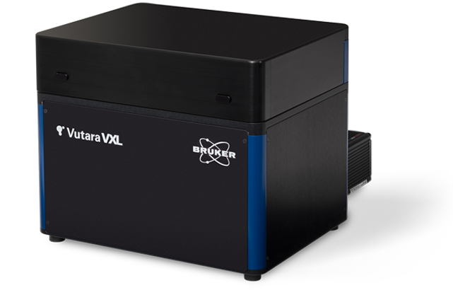
Vutara VXL
The Bruker Vutara VXL super-resolution microscope incorporates industry-leading single-molecule localization microscopy (SMLM) technology in a streamlined system with a compact footprint. The Vutara VXL system allows researchers to perform advanced research on DNA, RNA, and proteins including macromolecular complexes, super-structures, chromatin structures, and chromosomal substructures. This novel system also supports the study of functional relationships in genomes and various subcellular organelles and advanced spatial biology research in extracellular matrix structures, extracellular vesicles, virology, neuroscience, and live-cell imaging.
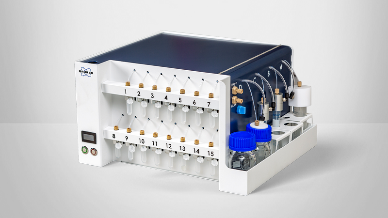
The Vutara VXL system incorporates a unique bi-plane detection technique to achieve 3D sub-diffraction resolution without any compromise in speed or sensitivity. Furthermore, when combined with Bruker's unique microscope fluidics unit, Vutara VXL enables multiplexed imaging for targeted, sub-micrometer multiomics in genomics, transcriptomics, and proteomics research.
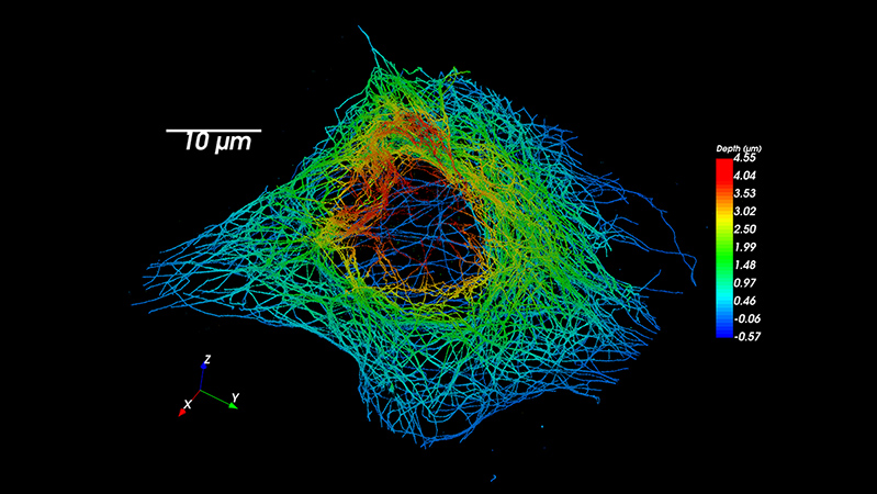
Super-Resolution Acquisitions in 3D
The Vutara VXL is equipped with proprietary bi-plane technology that enables you to gather 3D data with every acquisition. This technology allows you to easily perform a Z-stack series for thicker specimens and automatically localize and reconstruct the entire volume. With Vutara VXL, you can obtain comprehensive 3D data of your samples with ease.
Single-Molecule Imaging Beyond the Cover Slip
Vutara VXL is capable of imaging far from the surface of the coverslip, up to 100 µm deep, to accommodate a wide range of sample types. Thanks to the proprietary bi-plane technology, Vutara VXL with SRX software can perform single-molecule localization microscopy on more sample types than any other commercial single-molecule localization microscope available. Cultured cells, cell colonies, tissue sections, and entire model organisms are now accessible for single-molecule localization experiments.

Analyze Localizations with SRX Software

Vutara's Quantitative Localization Microscopy suite enhances your productivity and allows you to turn localizations into meaningful results. Vutara's SRX workflow-driven software guides users through the setup, calibration, imaging, processing, and analysis of the super-resolution single-molecule localization experiment. SRX boosts the value of the system by providing dedicated software that is designed specifically for SMLM. The SRX software combines real-time localization processing with advanced visualization and sophisticated quantitative analysis tools to let researchers quickly create publication-quality videos, images, and measurements.
SMLM Imaging Techniques on the Vutara VXL
Stochastic Optical Reconstruction Microscopy (STORM) and Direct Stochastic Optical Reconstruction Microscopy (dSTORM) enable localization of individual molecules using photoswitching or photo-activatable dyes that can allow high localization precisions — even in dense populations.
All SMLM methods rely on the selective on-off switching of fluorescent molecules. Classical STORM uses dye pairs (e.g., Cy3-Cy5), while dSTORM6 (direct STORM) requires only one fluorophore (e.g., Alexa647, Cy5) and a "switching buffer." Nevertheless, in both STORM and dSTORM, illumination with different wavelengths switches these fluorescent dyes on and off.
The advantage of STORM7 over PALM is that these dye molecules are highly efficient with large photon budgets, where the photo-activated proteins used in PALM emit a limited number of photons irreversibly photobleaching. As a result, STORM can achieve higher resolutions.


Photoactivated Localization Microscopy (PALM) enables localization of individual molecules within a specimen with high localization precision via the use of photoactivatable proteins. These fluorescence proteins are larger in size than the organic dyes and have lower photon budgets, but they allow endogenous labeling of proteins of interest.
Similar to STORM, PALM uses photo-activatable fluorescence proteins (e.g., paGFP, mEOS2), which can be easier than labeling with photoswitching dyes. These proteins are switched on with one laser wavelength and imaged and then bleached with another.
The primary difference between PALM and STORM -- to the advantage of PALM -- is that it allows protein labeling at the endogenous level. Moreover, because other SMLM techniques provide many localization events per molecule, PALM, which provides a single localization event per molecule, is often more applicable for experimental objectives related to stoichiometry/true moleculart counting.
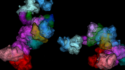
EXAMPLE APPLICATIONS OF THIS TECHNIQUE
- Exploration of how postsynaptic scaffolding proteins cluster on activity with 25 nm resolution8

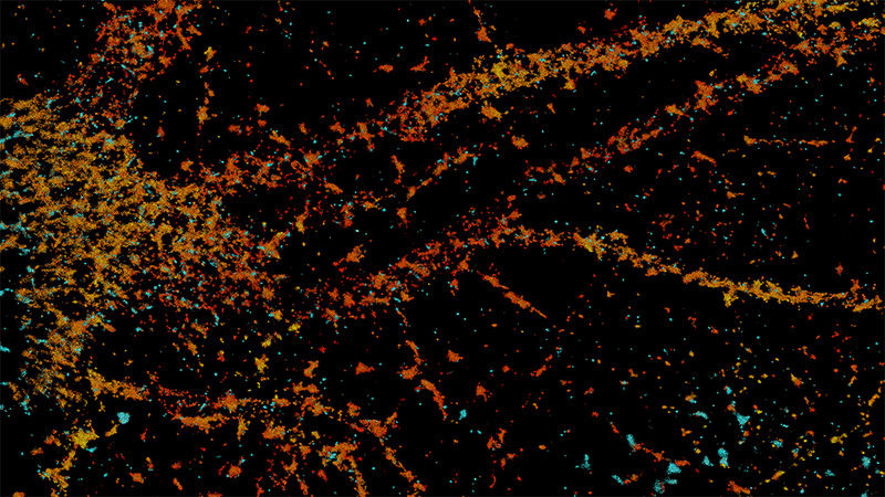
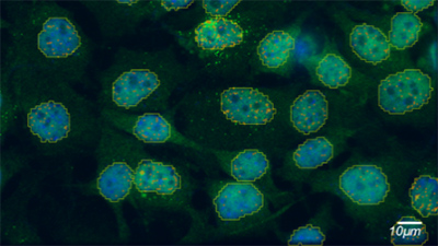
C. elegans as Model System for Synaptic Transmission.
Images courtesy of Sean Merrill and Rob Hobson from the Erik Jorgensen laboratory at the University of Utah (Utah, USA).

C. elegans as Model System for Synaptic Transmission.
The Jorgensen laboratory at the University of Utah (Salt Lake City, UT) uses the Bruker Vutara super-resolution platform to understand synaptic transmission in the worm C. elegans.
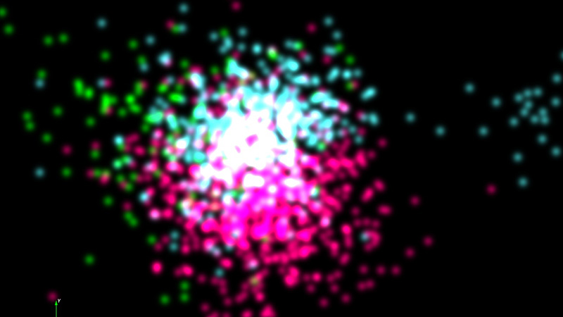
Point Accumulation for Imaging Nanoscale Topography (PAINT) allows for a potentially unlimited number of probes to be analyzed with high precision and resolution. It is also immune to photobleaching and overcomes the resolution limits of other SMLM techniques resulting from the photon budget of the chromophores used for labeling structures.
The other SMLM techniques require sequentially staining target biological structures and repeatedly imaging them to produce a composite image. In PAINT methods, fluorophores are no longer permanently bound to targets, but rather float in buffer solution. Instead of a laser, transitioned binding and immobilization of labels (e.g., Nile red) out of the buffer to the investigated structure provide the on-off mechanism. This allows imaging of an unlimited number of structures of interest in the same sample and makes PAINT methods easier and more efficient to implement than other SMLM methods.
While photobleaching diminishes the photon budget of the chromophores used for labeling structures in other SMLM techniques, PAINT10 methods are immune to photobleaching. As a result, PAINT methods can achieve a higher spatial resolution than other techniques.
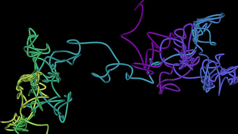
EXAMPLE APPLICATIONS OF THIS TECHNIQUE
- Precise colocalization studies of synaptic proteins9
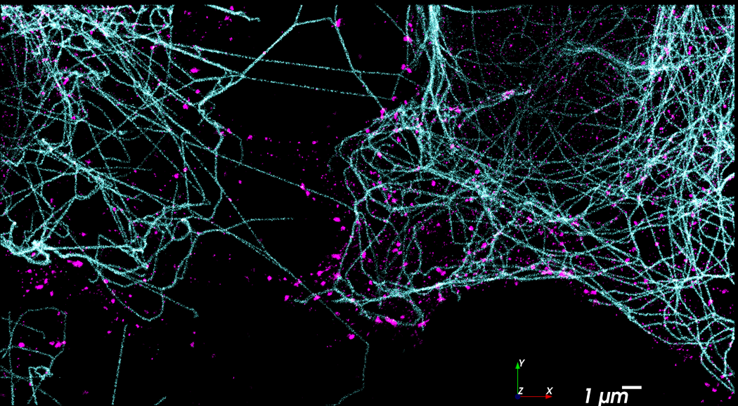
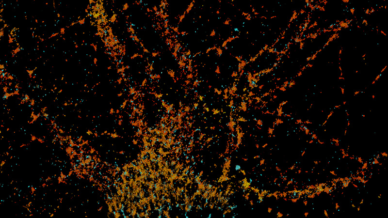
Images Automatically Collected Using the Vutara VXL with Fluidics
BS-C-1 labeled with anti-tubulin, actin, anti-tom20 and anti-clathrin. Orthogonal 2º DNA-PAINT antibodies were purchased from Massive-Photonics.com.


Single Particle Tracking (SPT) is the process of tracking moving particles in living cells. This allows for measurements of live cell dynamics such as the movement of particles within the cell, the connectivity of various compartments within the cell, or the production and location of newly created biomolecules. The tracked particles can be anything from genetically encoded fluorescent proteins to beads affixed to an antibody or nucleic acid sequence–in each case, the particles can be localized and tracked in 3D.
In single particle tracking, individual particles are tracked as they move around a living cell. The kind of tracked particles can vary. Single particle tracking was the first Single Molecule localization technique to be published, in a seminal work, a fluorescent bead was affixed to myosin, and its step-size determined. Beads can be affixed to biomolecules of interest either chemically or by interaction with e.g. an antibody. But many other single particle tracking techniques have been developed. In SPT-PALM, a portion of a population of proteins are photoconverted or photoactivated, and tracked until they bleach – this technique allows for tracking the dynamics of endogenous proteins with the cell. SPT is not limited to proteins – systems such as SP2, RNA Aptamers such as Peppers, or non-catalytic dCAS9 can be used to track the production, location and movement of RNAs, and dCAS9 or the LacI/Lac-O system the precise location of gene loci within chromatin as well. On the Vutara VXL, two or more classes of biomolecules can be tracked simultaneously in 3D with our dual camera accessory. The VXL is particularly well suited to single particle tracking as all particles within a roughly 1 micron Z section a 50x50 micron field of view can be localized and tracked natively in 3D at very high temporal resolution, and our powerful SRX software will handle the particle tracking for you.
EXAMPLE APPLICATIONS OF THIS TECHNIQUE
- Tracking the movement of RNAs down the axon
- Seeing if a protein of interest is actively localized or diffuses to its functional site
- Determining if different organelles or cellular compartments are connected or constrained
- Determining the rate of receptor pairing
- Measuring the rate of RNA production and correlate it to the position of a locus in the nucleus
Specifications
| Super-resolution localization microscopy (SMLM) | |
| Widefield microscopy |
|
| Excitation lasers (nominal laser power at diode) |
|
| Flat illumination |
|
| Multi-color acquisition |
|
| Multi-plane imaging |
|
| Camera |
|
| Objective |
|
| Field of View (FOV) |
|
| 3D SMLM method |
|
| SMLM resolution |
|
| Imaging depth |
|
| xy-stage |
|
| z-focus |
|
| Drift correction |
|
| Environmental control |
|
| Microfluidics unit for sequential labeling |
|
| Compact table-top design |
34 cm x 40 cm x 34 cm, (1.2 ft x 1.3 ft x 1.2 ft) 35 kg (78 lbs)
33 cm x 53 cm x 32 cm (1.0 ft x 1.7 ft x 1.0 ft) 30 kg (65 lbs)
53 cm x 35 cm x 14 cm (1.7 ft x 1.1 ft x 0.4 ft) 8 kg (17 lbs) |
| Operating in a typical laboratory environment |
|
| Vibration insulation included |
|
| Workflow-defined software for easy data acquisition |
STORM / dSTORM PALM PAINT Other blinking-based modalities
Chromatin tracing smFISH
Tracking of probe cycles and positions Support for an unlimited number of probes
|
| SMLM processing |
|
| Particle tracking |
|
| Fluidics Manager |
|
Drift correction |
|
| Statistical analysis tools |
Ripley’s K functions Pair correlation Nearest neighbors
DBSCAN OPTICS Delaunay Particle counts Volume and density calculations Determination of radius of gyration and sphericity Principal component analysis (PCA) Calculation of centroid
Pearson’s correlation coefficient Mander’s overlap coefficient Intensity correlation quotient Cross pair correlation Joint histogram STORM-RLA
Fourier ring correlation Labeling density resolution analysis Local resolution correlation mapping Nearest neighbors’ analysis |
| Chromatin tracing (optional) |
|
| Integrated visualization |
|
| Open data formats |
|
| Network attached storage (NAS) unit (optional) |
|
A Complete Software Solution, From Acquisition to Analysis
With SRX software and its Quantitative Localization Microscopy analysis suite, Vutara VXL can provide visual and quantitative information from biological samples. By localizing individual molecules, Vutara VXL can generate 3D images while simultaneously providing in-depth quantitative analysis tools.

Read What Our Customers Are Saying

With more than 15 years of experience in designing microscopes in academia and in industry, I am proud to contribute to the development of Vutara's super-resolution microscopes. With biplane 3D detection and fast sCMOS imaging, Vutara has the most advanced super-resolution microscope on the market.
Joerg Bewersdorf, Ph.D., Yale University

After scanning the market for super-resolution microscopes and personally visiting and testing most commercially available systems with our own samples, I can say that I am most impressed with Vutara's SR-350. In particular, the user friendliness of their imaging software as well as the 3D capability in super-resolution mode impressed me.
Vutara has an excellent support team and staff as a whole making our transition to super-resolution a well-supported experience. If you're looking to advance your research by super resolution microscopy, I can confidently say that Vutara's systems are an excellent choice.
Thomas Stroh, Ph.D., Core Facility Director, McGill University

The volume of data you can acquire on a super-resolution system is incredible when comparing electron microscopy especially considering the sample preparation.
Peter McPherson, Ph.D., McGill University

Having published with a Vutara super-resolution system, I can confidently say that they offer one of the most advanced super-resolution microscopes available. Their attention to detail and ongoing close collaboration makes them one of my preferred microscopy vendors.
Brigitte Ritter, Ph.D., Boston University