Guide to FT-IR Imaging
What is an FT-IR image?
An FT-IR image is a type of chemical image created by an FT-IR microscope. Chemical images combine the digital topographical data of a sample obtained with a microscope with chemical information obtained through spectroscopy. This allows us to visualize the chemical composition of a sample.
FT-IR imaging is an exceptionally useful technique to analyze the distribution of compounds in a sample or to help understand the structure of a sample. This technique has broad ranging applications whether that is in research, medicine, quality control, or failure analysis.
How FT-IR images are created
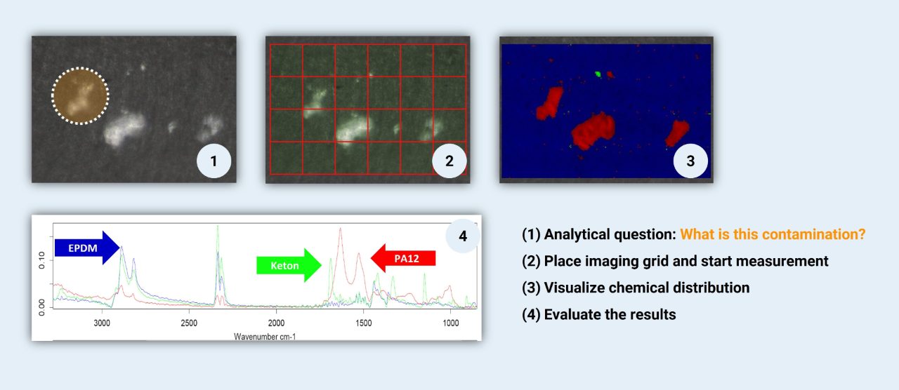
The fundamental technique behind FT-IR imaging is FT-IR spectroscopy, a chemical analysis technique that detects how different compounds interact with infrared light. Different compounds have unique interactions with infrared light, so the FT-IR spectrum that is created from FT-IR spectroscopy acts as a “chemical fingerprint” to identify components in a sample.
The FT-IR microscopes used to create FT-IR images combine a traditional microscope with FT-IR spectroscopy. The first step to create a chemical image is to first obtain a digital image of the sample using the microscope. A region of the sample is then selected for analysis. Then, the IR data is collected across that entire region of interest such that every point on the sample will correspond to an IR spectrum.
Depending on the sample there are several measurement techniques that can be used to collect that data: transmission, reflection, or attenuated total reflectance (ATR). Any measurement technique can be used to analyze the sample with the FT-IR microscope.
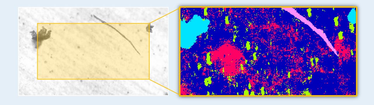
Now that each point in the region of interest corresponds to an FT-IR spectrum, all those spectra can be analyzed to see what compound are present in the sample. This can be done manually or automatically with computer softwareq or even artificial intelligence algorithms. After analysis, each compound is assigned a different color. Then, every pixel on the image can be colored according to which compound is present at that point to create the chemical image (3).
Detectors for IR Imaging
To create chemical images, IR light must be detected after interacting with the sample to create the spectrum. There are several detector options for FT-IR microscopes. The typical choice is a single-element detector, which can obtain an IR spectrum at one point on the sample. Of course, you can now set many points in close proximity across the sample to create an image.
However, analyzing an entire region of the sample this way would take a very long time which is only attractive if labor cost ist low. If time is money, special imaging detectors are used in FT-IR imaging instead. There are two types of imaging detectors: line array or focal plane array (FPA) detectors.
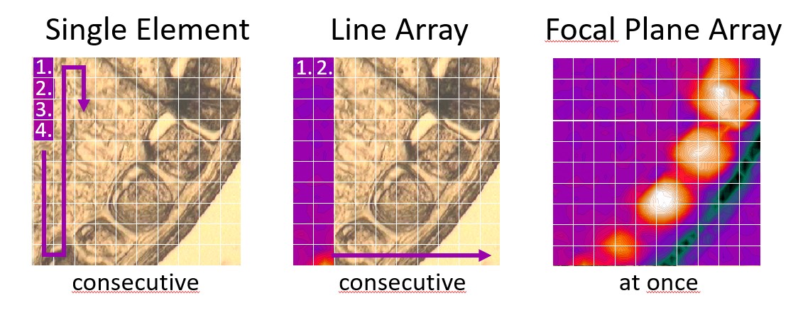
Line array detectors are a somewhat cheaper, pseudo-imaging solution. These detectors are made of several single-element detectors arranged in a line, so multiple spectra can be obtained simultaneously. The sample is then scanned line-by-line, and the lines of spectra are stitched together to create the chemical image.
FPA detectors are the state-of-the-art detector technology for FT-IR imaging. These detectors are made of an array of IR detectors arranged in a square (e.g. 64 x 64 detectors). With that many detectors, thousands of spectra can be captured in a single shot, almost like a digital camera. If a thermal source is used, these detectors are liquid nitrogen cooled, if an IR laser is used as a source, uncooled FPA microbolometers are used.
FPA detectors far exceed the capabilities of line array and single element detectors. Chemical images of even large areas can be generated quickly, with excellent resolution so means the highest definition images can be created in the shortest amount of time.
Applications of IR Imaging
FT-IR and IR laser imaging are generally applicable whenever there is a need to visualize the distribution of chemical compounds in a sample. This is commonly used in industry to check products for uniformity or contamination.
Checking a product for uniformity is particularly important in the pharmaceutical industry. Various drugs that come in tablet form ought to be completely uniform, so that there are no “hotspots” of the active ingredient. Afterall, many tablets can be broken in half to split the dose, so it would be problematic if the active ingredient was only in half of tablet. IR imaging can quickly scan the entire tablet and the chemical image is an easy way to see the distribution of the active ingredient to ensure it is evenly distributed.
Examining finished products for contaminants is an important aspect of quality control in many industries. IR imaging has the advantage here since it can quickly scan large work pieces. For example, large silicon wafers produced in the semiconductor industry can be imaged in just a few minutes with an FPA detector to see if there are any impurities in the wafer. Even after the wafer has had electronics soldered to it, IR microscopy can still be used for failure analysis on even the hard-to-reach areas of the chip.
Beyond these standard quality control and failure analysis applications, creating highly detailed chemical images is useful in many fields to visualize and characterize the structure of a sample, from forensics, to paleontology, to art restoration, to medicine.
One particularly exciting application of IR imaging is tissue imaging, especially using IR lasers. With an FPA detector, even large sections of a tissue sample can be imaged in just a few minutes. These images can clearly show the distribution of carbohydrates, proteins, and lipids in the tissue. This could be extremely beneficial to the medical field, where patient tissue samples can be rapidly screened for disease or cancerous cells. IR imaging is much faster than other methods to screen tissues, so this technique has the potential to speed up the process disease and cancer detection significantly, providing quick answers to patients.
Frequently asked question about FT-IR imaging
1. What is chemical imaging?
Chemical imaging is a method for spatially resolving the chemical properties of a sample in 2D or 3D images. With this technique it is possible to obtain information about the material properties, the structure and the origin of the examined samples.
2. What is FT-IR imaging?
FT-IR imaging is one way to create said spatially resolved chemical images. Each pixel of these images consists of a whole IR spectrum. By interpreting the individual spectra, interesting sample regions can be detected and evaluated.
3. How do you create FT-IR images?
Common methods are sequential single point or line array measurements, as well as the direct acquisition of 2D images by a focal-plane array (FPA) detector. While FPA detectors offer the superior solution, highly automated single-point measurements are an economical alternative.
4. How does an FPA detector work?
The principle of an FPA detector is analogous to that of a digital camera. Instead of visible light, however, a defined array of pixels is illuminated by infrared light, with each detector pixel recording an independent, spatially resolved IR spectrum.
5. Do FPA detectors require apertures?
No, an FPA detector does not require any apertures. Each pixel of the detector functions as an aperture and thus records a spatially IR information directly. This allows much faster and higher resolution measurements compared other detector techniques.
6. Is it possible to adjust the spatial resolution of an FPA?
The spatial resolution of an FPA detector depends on the size of the individual detector pixels. However, adjacent pixels can be combined to form a "larger pixel" and thus the spatial resolution is reduced, also improving spectral quality.
7. Are there different FPA sizes?
FPA detectors are available in different array sizes. Size should be selected according to the optical system (microscope). For example, the LUMOS II is optimized for a 32x32 pixel array, while the HYPERION 3000 is designed for a 64x64 or 128x128 pixel arrays. With the latter it is possible to record an impressive number of more than 16,000 spatially resolved spectra in one scan.
8. Is a larger FPA better?
No, because the size of the FPA detector depends exclusively on the optimal illumination provided by the microscope. A homogeneous illumination of the detector array is important to ensure a consistently high spectral sensitivity both in the center and at the edges of the detector.
9. When does a larger FPA have advantages?
The larger the FPA detector area, the more spectra are recorded simultaneously. Since the spatial resolution is independent of the array size, this means that a 128x128 FPA detector covers an area 16 times larger than a 32x32 detector array in a single measurement.
10. Can FPA be combined with any measurement technique?
Yes they can. FPA detectors offer advantages in transmission, reflection and attenuated total reflection (ATR). Especially when used with ATR technology, this type of detector achieves an exceptionally high spatial resolution.
11. Why is the resolution of FPA measurements in ATR increased?
The combination of a high refractive solid-state lens (germanium ATR crystal) and an "aperture-free" FPA detector increases spatial resolution by a factor of 4 compared to transmission measurements. This effect is also called an immersion lens.
12. Are FPA measurements applicable to all samples?
Since FPA measurements can be combined with all measurement techniques, in principle all types of samples can be analyzed this way. Gases, liquids and other volatile substances cannot be analyzed microscopically due to their kinetic properties.
13. What are typical applications of an FPA?
Typical applications can be found in all areas of industry and research. Starting with the analysis of microplastics, particles and contaminations over the characterization of complex chemical structures, such as biological tissue, pharmaceutical products up to multilayer laminates and lacquers. In short, this detector technology is used wherever very high spatial resolution and the analysis of large sample areas are indispensable.