
Episode 6: The Role of Mechanical Forces in Health and Disease
Episode 6: The Role of Mechanical Forces in Health and Disease
In this episode, we had the pleasure of speaking to Prof. Elisabeth Fischer-Friedrich from the Cluster of Excellence 'Physics of Life' at the Technical University Dresden, Germany, about The Role of Mechanical Forces in Health and Disease.
Prof. Fischer-Friedrich talks about her interdisciplinary research investigating how stress and mechanical properties influence biological function and delves into the interplay between intracellular forces, deformation, and dynamic mechanical properties.
TO LEARN MORE:
The Role of Mechanical Forces in Health and Disease
When we think about biology, we often think about the DNA double helix, proteins, or high-resolution images of cells. What is less prominent in our minds, but equally fundamental, are the mechanical forces that act on and in cells. These forces shape tissues during development, drive cell migration, influence disease progression, and play many more roles [1], [2].
To understand the forces and biophysics of cells, researchers combine biology, physics, and advanced microscopy to uncover how living matter behaves under stress. In Episode 6 of the Conversations on AFM podcast, we speak with Prof. Dr. Elisabeth Fischer-Friedrich about her research and how she investigates cells and tissues responding to forces, ultimately exploring how this affects health and disease. Prof. Dr. Fischer-Friedrich is a Heisenberg Professor at the Physics of Life Cluster of Excellence at TU Dresden.
Why Mechanics Matters in Biology
Mechanical forces constantly act on cells as they are being pushed, pulled, and squeezed, both by their environment and by the forces they generate themselves. According to Prof. Fischer-Friedrich, these forces directly affect the material properties of living matter [3].
Cells are considered a viscoelastic material [4], [5] and can stiffen or soften in response to stress. If they are soft, they easily deform; if stiff, they resist external pressure. Much of this behaviour comes from the actomyosin network, a dynamic scaffold beneath the cell membrane that generates active forces; similar to muscles in our body, but on a (sub-)cellular scale.
Importantly, cells are not static. They can regulate their own stiffness by altering actomyosin activity. High activity increases stiffness, while lower activity relaxes the cell. Additionally, molecular turnover allows cells to behave like solids on short timescales but like fluids over longer periods. Functionally integrating this multi-level complexity is central to tissue growth, wound healing, and morphogenesis.
Measuring the Mechanics of Cells
But how can scientists measure something like cell stiffness?
This is where atomic force microscopy (AFM) becomes indispensable.
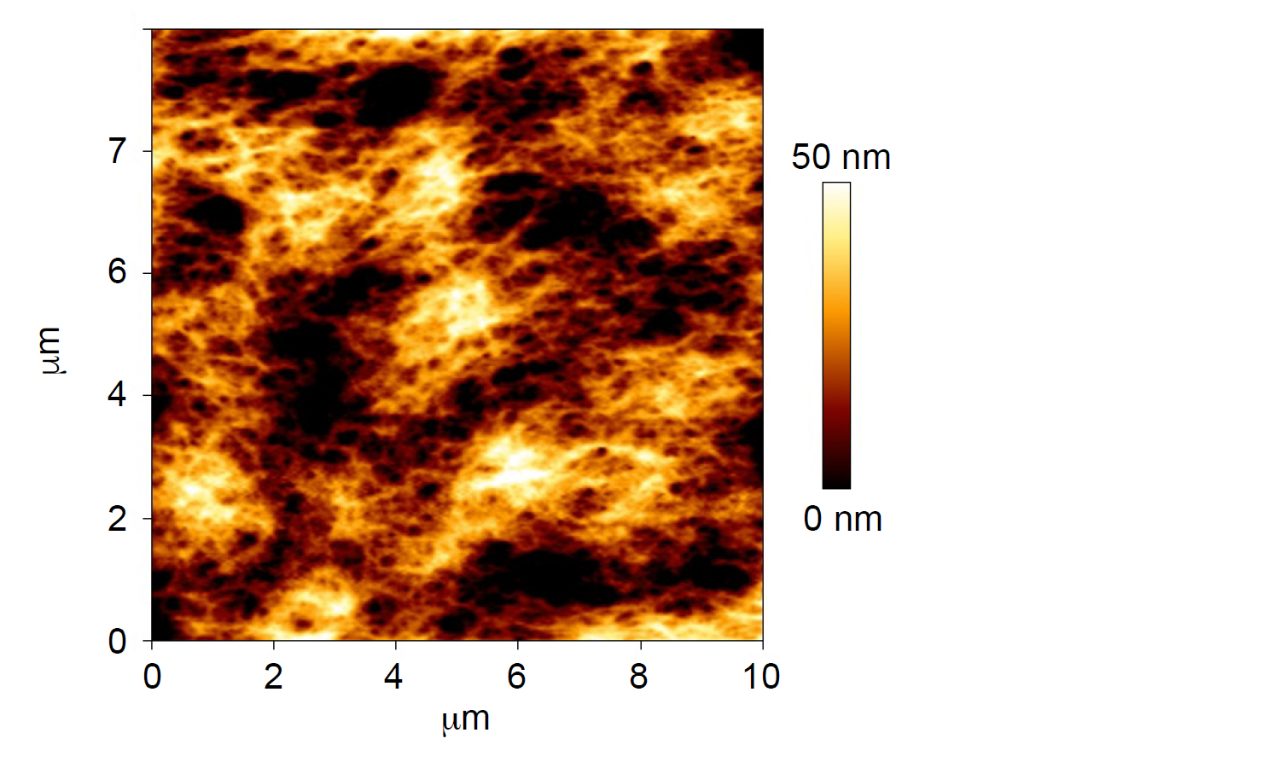
AFM is a technique that belongs to the broader family of scanning probe microscopies. Unlike optical microscopes, which use light, AFM “feels” the surface of a sample with a nanoscale tip attached to a cantilever. In biological research, applications of AFM range from imaging the surface of cell membranes, probing the elasticity of tissues, to measuring the unfolding of individual proteins.
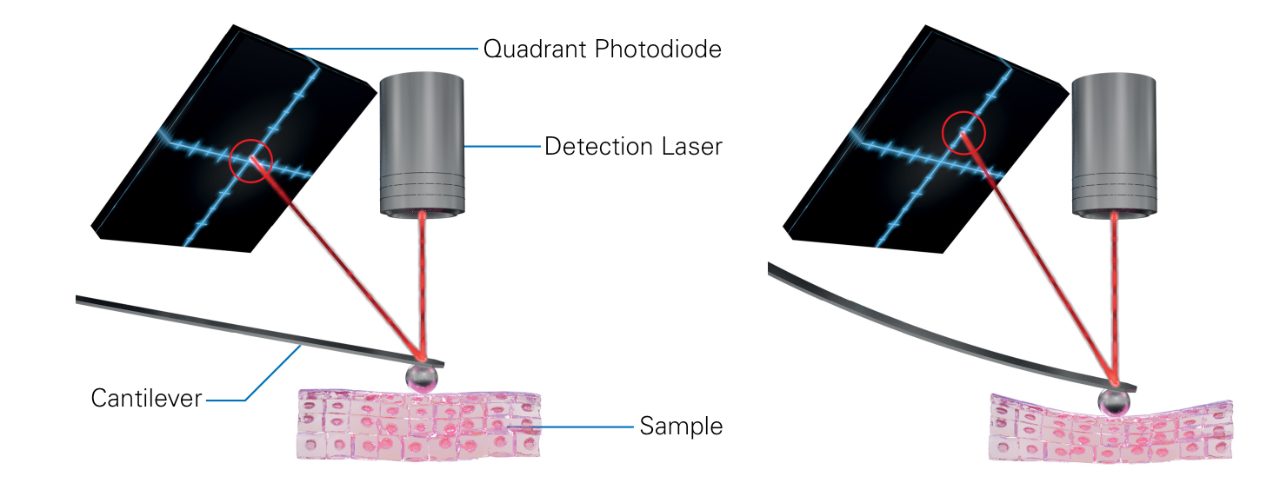
By applying forces and recording the cantilever’s deflection, AFM can create detailed topographical maps and measure how soft or stiff a material is. In AFM indentation, the Hertz model of contact mechanics is most commonly applied, assuming the sample behaves as an isotropic, uniform material characterized by a defined Young’s modulus and Poisson’s ratio.
The Basement Membrane: A Tissue’s Mechanical Scaffold
One area where these methods shine is the study of the basement membrane. This thin, specialized extracellular matrix - that supports epithelial and endothelial tissues - provides both structural support and signalling cues.
Prof. Dr. Fischer-Friedrich’s lab, in collaboration with Prof. Dr. Christian Dahmann at TU Dresden, applied AFM to probe the basement membrane in Drosophila tissues. Interestingly, unlike the rapidly turning over actin cortex inside cells [6] basement membranes are remarkably stable, with turnover times ranging from hours in embryos to years in adult humans. This stability allows basement membranes to behave like elastic solids, capable of bearing long-term stresses [2], [7].
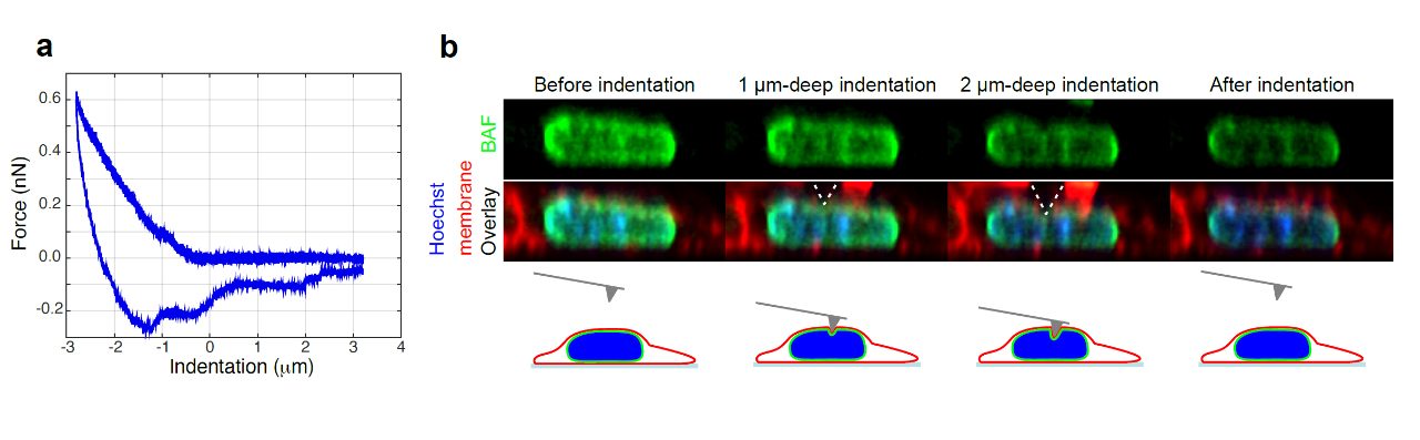
Using AFM indentation, the Fischer-Friedrich team discovered that basement membranes are under pre-existing mechanical tension, much like a trampoline stretched tight. This finding reveals that tissues are not passive scaffolds; they are under constant internal stress that shapes their behaviour during development, ageing, and disease.
Biophysics in Health and Disease
Cells continually adjust their mechanical properties to meet the demands of their environment. This is the case in health and disease, such as developmental tissue folding or cancer metastasis.
In cancer, cellular biomechanics take on a particularly critical role. Prof. Fischer-Friedrich’s group has shown that as cells prepare to divide, contractility in the actin cortex increases, producing the tension needed for mitotic rounding [8]. This change allows cells to adopt a spherical shape and complete division even in the crowded conditions of a tumour.
Her team has also explored mechanical changes during epithelial–mesenchymal transition (EMT), a key step in metastasis. Here, cells become softer in interphase, aiding migration, but stiffen again during mitosis, preserving their ability to proliferate [9], [10].
Towards the Physics of Life
For biophysicists like Prof. Fischer-Friedrich, AFM is more than an imaging tool; it is a quantitative instrument that reveals how the fundamental physics of biomechanical force and material properties underpin biology. By combining AFM, mathematical modelling, and creative experimental design, the Fischer-Friedrich lab helps to gain insights into the physics of life.
You can also download our ressource collection to learn more about AFM technologies and their application.
References
- V. Tsata and D. Beis, “In Full Force. Mechanotransduction and Morphogenesis during Homeostasis and Tissue Regeneration,” Journal of Cardiovascular Development and Disease, vol. 7, no. 4, p. 40, Dec. 2020, doi: 10.3390/jcdd7040040.
- K. Y. G. Santillán, C. Jantzen, C. Dahmann, and E. Fischer-Friedrich, “Epithelial Folding Through Local Degradation of an Elastic Basement Membrane Plate,” Advanced Physics Research, vol. 3, no. 12, p. 2400062, 2024, doi: 10.1002/apxr.202400062.
- E. Fischer-Friedrich, “Active Prestress Leads to an Apparent Stiffening of Cells through Geometrical Effects,” Biophysical Journal, vol. 114, no. 2, pp. 419–424, Jan. 2018, doi: 10.1016/j.bpj.2017.11.014.
- K. E. Kasza et al., “The cell as a material,” Current Opinion in Cell Biology, vol. 19, no. 1, pp. 101–107, Feb. 2007, doi: 10.1016/j.ceb.2006.12.002.
- M. Mokbel, K. Hosseini, S. Aland, and E. Fischer-Friedrich, “The Poisson Ratio of the Cellular Actin Cortex Is Frequency Dependent,” Biophysical Journal, vol. 118, no. 8, pp. 1968–1976, Apr. 2020, doi: 10.1016/j.bpj.2020.03.002.
- E. Fischer-Friedrich, Y. Toyoda, C. J. Cattin, D. J. Müller, A. A. Hyman, and F. Jülicher, “Rheology of the Active Cell Cortex in Mitosis,” Biophys J, vol. 111, no. 3, pp. 589–600, Aug. 2016, doi: 10.1016/j.bpj.2016.06.008.
- K. Y. Guerra Santillán, C. Dahmann, and E. Fischer-Friedrich, “Elastic Contractile Stress in the Basement Membrane Generates Basal Tension in Epithelia,” PRX Life, vol. 2, no. 1, p. 013004, Jan. 2024, doi: 10.1103/PRXLife.2.013004.
- E. Fischer-Friedrich, A. A. Hyman, F. Jülicher, D. J. Müller, and J. Helenius, “Quantification of surface tension and internal pressure generated by single mitotic cells,” Sci Rep, vol. 4, no. 1, p. 6213, Aug. 2014, doi: 10.1038/srep06213.
- K. Hosseini, A. Frenzel, and E. Fischer-Friedrich, “EMT changes actin cortex rheology in a cell-cycle-dependent manner,” Biophys J, vol. 120, no. 16, pp. 3516–3526, Aug. 2021, doi: 10.1016/j.bpj.2021.05.006.
- K. Hosseini, A. Taubenberger, C. Werner, and E. Fischer-Friedrich, “EMT-Induced Cell-Mechanical Changes Enhance Mitotic Rounding Strength,” Advanced Science, vol. 7, no. 19, p. 2001276, 2020, doi: 10.1002/advs.202001276.
Interested in learning more about this topic?
You can find detailed applications and technical notes, expert-led webinars, and on-demand instrument and measurement demonstrations in our online resource library. Get instant, full-length access to all resources related to this podcast using the form below.
This resource collection includes:
- 2 full-length e-books
- 2 full length on-demand webinar recordings
- 2 real-time instrument demonstrations


