Unlike neurons, the glial cells known as astrocytes show minimal electrical activity and use calcium to mediate intracellular functions. Therefore, although their important role in mammalian sleep was postulated their interplay with neurons during sleep is not fully understood.
The authors studied astroglial calcium dynamics during sleep-wake cycle and role of astroglia in homeostasis of sleep which is understood as sleep intensity and/or sleep duration that depends on prior time spent awake.
The astroglial calcium dynamics were monitored in the frontal cortex in mice. This is because frontal cortex shows greatest dynamic range for non-rapid eye movement sleep (NREMS), slow wave activity (SWA), a canonical index of mammalian sleep need. In parallel to recording calcium changes in astroglia, the electroencephalographic (EEG) and electromyographic (EMG) activity was recorded in un-anesthetized, freely behaving mice.
First, the authors showed that astroglia calcium signals were changing with vigilance state. At this stage of their work they used head-mounted, miniature microscope.
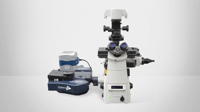
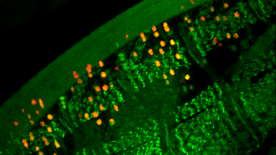
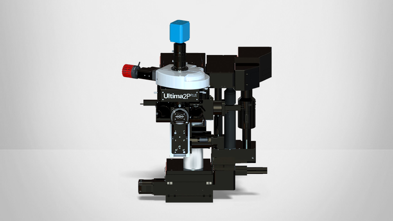
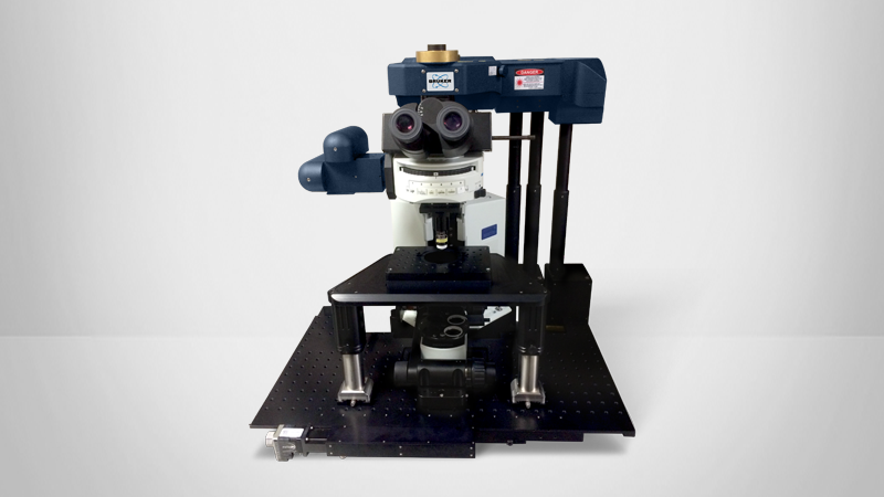
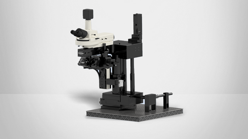
Next, they investigated whether changes in calcium signals were uniform throughout the astrocyte or compartmentalized in the soma versus processes. Because the signal captured by the miniature microscope includes both somatic and process activity, they used Ultima IV (intravital, two-photon microscope, Bruker Corp.) to more precisely examine how astroglial calcium signals change in these subcellular regions in frontal cortex in un-anesthetized mice across vigilance states. They found that during wake, NREMS, and REMS, astroglial processes showed greater frequency of single calcium events compared to somata.
The authors then tested whether changes in astroglial calcium encode changes in sleep need. They recorded calcium changes on Ultima IV in astrocytes of sleep deprived mice. They found that sleep deprivation induced changes in astroglial calcium during NREMS and primarily increases in processes.
Sleep is accompanied by changes in the synchrony of neuronal networks. Using the miniature microscope, the authors found that astroglia were highly synchronized in all brain states, but this synchrony was greatest during wake and lowest in sleep. Sleep deprivation increased synchronized neuronal activity in NREMS whereas sleep deprivation reduced synchronized astroglial activity in NREMS but increased it in REMS. This means that astroglial calcium signals, at the network level, became less synchronous during the recovery from sleep-deprivation.
Finally, the authors deleted STIM1 in astrocytes. STIM1-mediated store-operated calcium entry (SOCE) is an essential mechanism by which the intracellular calcium concentration is elevated. Inhibiting STIM1 impairs SOCE in astrocytes. Interestingly, diurnal/nocturnal patterns of running wheel activity, cage activity, and core body temperature did not differ between wild type and knock-out mice. After sleep deprivation, however, the response to the lack of sleep was blunted in knock-out mice as measured by NREM SWA.
These findings demonstrate that sleep in not only accompanied by widespread activity changes in neurons but also by changes in glial cells. The further results suggest that astroglial calcium signaling is part of the mammalian sleep homeostasis. Furthermore, this study highlights new players to take into consideration for sleep researchers.