Ultima 2Pplus
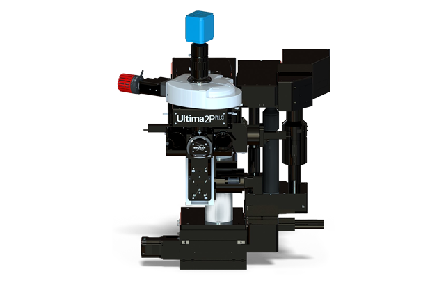
Ultima 2Pplus
With new advances in its electronics architecture (xCore), field of view, sensitivity, wavelength, and sample accommodation, the Ultima 2Pplus delivers an ideal combination of flexibility, resolution, imaging depth and speed, allowing users to perform simultaneous imaging, stimulation and electrophysiology protocols with greater efficiency and effectivity. The system is designed specifically for intravital imaging, with fully motorized control of the objective X-Y-Z position, as well as two axes of rotation for precise imaging orientation. A second scan path enables simultaneous imaging and photoactivation. The system’s optimized optical train confers exceptional performance to the very edges of the large field.
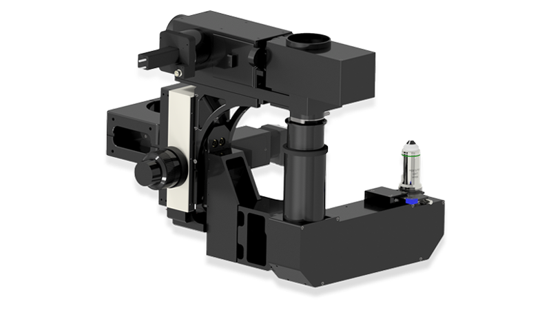
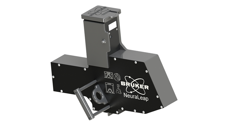
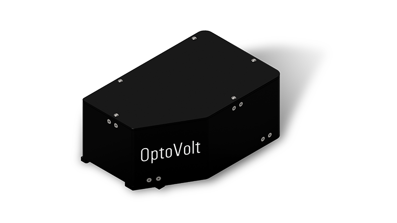
Add-on modules enhance the capabilities of the microscope: NeuraLight 3D Ultra offers the most advanced 3D holographic workstation for simultaneous all-optical stimulation and imaging; NeuraLeap is a groundbreaking Digital Micromirror Device that enables rapid imaging across a range of depths with greater temporal resolution; the OptoVolt module delivers unprecedented kilohertz imaging rates to enable voltage imaging neural research; the xView exchangeable lens set extends the field of view (FOV) by more than 2.5x the standard area; and the new Inverter module allows operators to easily transform the system into an inverted microscope without losing the quality and performance of the standard upright configuration.
Engineered for High-Performance Imaging
The Ultima 2Pplus redefines multiphoton imaging with a larger field of view, enhanced sensitivity, and deeper imaging capability, delivering high-quality data without trade-offs. Designed specifically for intravital imaging, it brings together advanced scanning, precise sample control, and an optimized optical path for deep, wide-field imaging in living systems.
For researchers, this translates to faster experiments, improved image clarity at depth, and the ability to observe and manipulate neural activity across larger, more complex biological networks.
Largest Field of View and Superior Field Uniformity
The Ultima 2Pplus field of view is over 50% larger to facilitate modern optogenetics experiments, which require simultaneous imaging and photostimulation of distant regions in a sample. A new, custom optical design preserves spatial and temporal resolution throughout the entire field, which is essential for researchers to be able to continue their work without changing experimental designs.
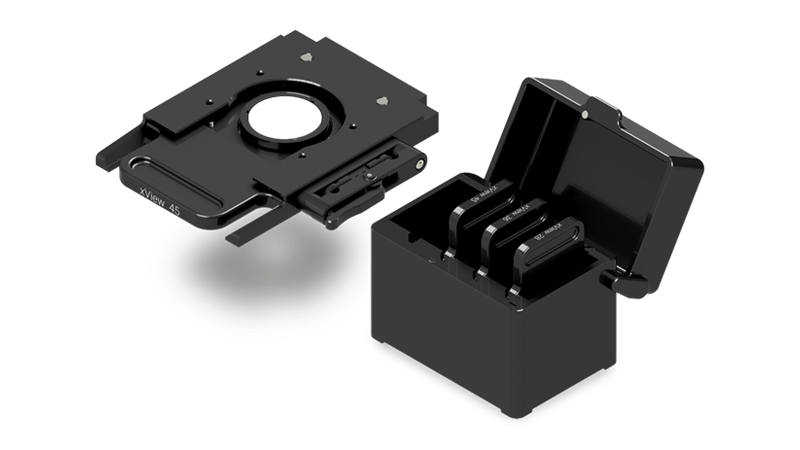
Additionally, Bruker’s xView Module delivers a breakthrough in large-scale, high-resolution imaging: this exchangeable lens set extends the field of view (FOV) by more than 2.5x the standard area, enabling researchers to visualize neural activity without sacrificing speed or sensitivity.


High-Sensitivity Deep Imaging
The Ultima 2Pplus light collection path ensures detection of almost all photons scattered from dim, thick and opaque biological samples. The superior collection efficiency is provided with 2-inch or larger optics throughout the emission path up to the sensitive close-proximity detectors.
As two-photon microscopy has become well adopted, emerging three-photon microscopy offers the potential to image even deeper into samples. Since further infrared wavelengths are scattered even less, three-photon excitation has the promise to be able to image several millimeters into the brain. Ultima 2Pplus already uniquely incorporates this capability to take your research to new levels of potential.
Perfected Photostimulation and Imaging
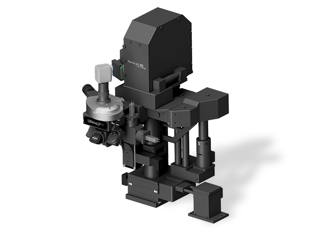
The Ultima 2Pplus empowers sophisticated optogenetics experiments with Bruker's new Neuralight 3D Ultra Spatial Light Modulator option. Large populations of targets can be stimulated simultaneously in multiple focal planes with computer-generated holograms. During 3D holographic experiments, targets at different depths are imaged with a custom-designed, optically-corrected ETL focusing module that allows for independent Z-positioning of the imaging plane relative to photostimulation.
Read a recent article “Cortico-cortical feedback engages active dendrites in visual cortex” published in Nature from Michael Häusser’s Lab. Bruker's two-photon calcium imaging, eletrotunable lens (ETL), and spatial light modulator (SLM) were used for long-range all-optical connectivity mapping and investigating feedback in the mouse brain.
Michael Hausser’s lab also achieved outstanding results with Bruker’s OptoVolt Module on the Ultima 2Pplus. They were able to precisely study the spatiotemporal relationships of hippocampal CA1 neurons with OptoVolt in 8x mode.
Other add-on modules are available to further enhance the capabilities of the microscope.

Optical stimulation of circled cells with Neuralight 3D. 3D hologram of points was created which was then spirally scanned over neuronal cell bodies.
Data courtesy of Adam Packer, Lloyd Russell, Henry Dalgleish, Michael Häusser’s lab, UCL, London.

OptoVolt 8x mode (550Hz) showing two-photon imaging of Hippocampal CA1 neurons expressing the voltage indicator FORCE1s, playing at 0.1x speed. Delta F/F in color with anatomical overlay in gray.
Data courtesy of Qiyuan Liang of Michael Häusser’s lab (Hong Kong University), and Francois St-Pierre (Baylor College of Medicine).
Ultima 2Pplus Data Gallery


Select Ultima 2Pplus Specifications
| SCAN HEAD | |
| Scanning Method | Matched pair of 6 mm Cambridge galvanometers with raster and spiral scanning capabilities |
| Field of View | ~1.375 mm x 1.375 mm with 16x objective (≤28 mm FN) |
| Scan Speed | Raster scan: 1.65 fps at 512 x 512, >12 fps at 64 |
| Scan Customization | User-definable straight, freehand and circular (infinite) linescan with included software; User-definable pixels/line and lines/scan from 1 to 2048; ≤120x scan zoom; 360° of scan rotation; Point scan |
| Uncaging Option | Second set of matched 3 mm or 6 mm Cambridge galvanometers in same scan head provide high-precision visible or multiphoton laser sample photomanipulation |
| High-Speed Imaging Option | 8 kHz resonant galvanometer; ~30 fps at 512 x 512, >1300 fps at 512 x 8 region of interest |
| DETECTORS | |
| Reflected Non-Descanned | 1 to 4 hand-picked Hamamatsu Multi-Alkali PMTs; Upgradeable to high-sensitivity Hamamatsu GaAsP PMTs |
| Transmitted Non-Descanned | 1 or 2 hand-picked Hamamatsu Multi-Alkali PMTs; Upgradeable to high-sensitivity Hamamatsu GaAsP PMTs |
| Dodt | Single Hamamatsu PMT for DIC-like image collection |
| Transmitted | Single Hamamatsu PMT for transmitted light image collection |
| Camera | Standard C-mount camera port built into scan head |
| OPTICAL INPUTS | |
| Multiphoton Laser | Optimized for multiphoton laser input from 690 to 1700 nm |
| Epifluorescence | High-Powered LED in epifluorescence turret |
| Visible Laser | Fibered laser inputs for visible laser introduction |
| Ultima Laser Rating | Class 1 (Contains Class 3b and Class 4 lasers) with light box |
| Helios Laser Rating | Class 3b |
| LED | Full-field photoactivation with LED module |
| MOTOR CONTROL | |
| Bruker X, Y Stage | Variable height with ~15 cm X and ~7.5 cm Y movement and ~0.3 μm step size |
| Bruker X, Y Microscope Base | Fine and coarse-movement platform for scope with ~35 mm travel and 0.1 μm step size |
| Bruker Z-Focus | Range of 30 mm with ~0.2 μm step size |
| Bruker Z-Piezo | Travel range ≤1000 μm with 0.05 μm step size |
| Bruker Orbital Nosepiece | Motorized control of objective angle and rotation |
| SOFTWARE | |
| Prairie View Imaging | Turnkey intuitive and customizable operation for imaging |
| Z-Series | Easy creation of depth stacks with user-customizable slice number, step size and laser power |
| T-Series | Easy creation of complex series involving Z-Series and triggered images |
| Stage Montage | Atlas Imaging simplifies setup and optimizes acquisition of 2D and 3D stage montages |
| Peripherals Integration | Wavelength and power control available for multiphoton and visible laser launches |
| Photoactivation | User-defined points and regions with synchronized laser modulation |
| Regions of Interest | User-defined regions for faster scanning capabilities |
| Brightness Over Time | Intensity mapping and plotting for user-defined regions over time |
| Voltage Inputs/Outputs | Signal inputs and outputs for electrophysiological experiments, stimulus control, and synchronization with external devices |
Hear What Our Customers Have to Say

We have the 2Pplus - the scope is well-designed and the software easy to use. Their scopes are very flexible in design if you wanted to add additional optics, components, or software functionality. Bruker shines with respect to customer service… they have several great service experts who usually respond within a day or two with insightful solutions. Further, the support is highly personalized; not before long after purchasing the microscope, we were paired with specialists who intimately familiarized themselves with our applications.
Charles Zhou, Ph.D., UW Center in Neurobiology of Addiction, Pain, and Emotion

We appreciate the modular nature of the Ultima hardware and software. The systems now available range from basic imaging systems to the dual-laser photo-stimulation multi-mode imaging workstation. We have continued updating many of our 14 machines, including our ten year old systems. The Team Viewer connection and support is a huge asset for novice owners and for significant software changes.
David Wokosin, Ph.D., Northwestern University