Ultima In Vitro
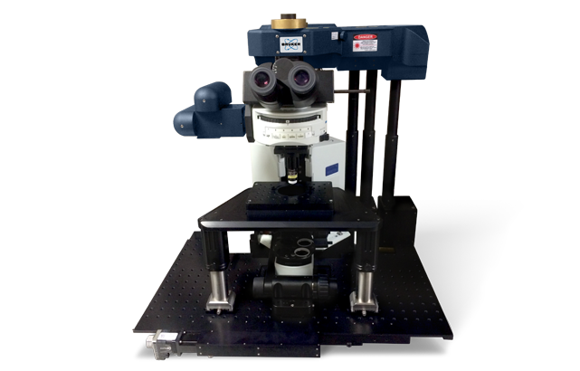
Ultima In Vitro
The Ultima In Vitro Multiphoton Microscope System is the platform on which leading scientists have conducted breakthrough experiments in brain-slice studies related to neural signaling and neural network mapping. Ultima In Vitro provides simultaneous imaging and electrophysiology, plus uncaging and photostimulation for the complete stimulus and recording environment required for research by today’s neuroscientist.
Flexible Architecture to Meet Varying Requirements
Ultima In Vitro is the ultimate “slice rig," having provided the platform on which leading scientists have conducted breakthrough experiments in brain slices related to neural signaling and neural networks for more than ten years. The system provides the flexibility for the most demanding protocols, and is available on upright or inverted optical microscopes. Features include:
- Secondary scan path that enables simultaneous imaging and photostimulation
- Visible, UV, and IR inputs for photostimulation
- Sub-micron positioning control for point photoactivation
- Sub-millisecond spirals for somal actiavtion
- Synchronization and triggering to enable simultaneous imaging, photostimulation, and electrophysiology
- Integrated electrophysiology control and recording with up to 16 analog outputs/inputs

High-Resolution Multifield Imaging

The Atlas Imaging module in Prairie View software simplifies setup of 2D and 3D automated montages. X,Y boundaries are easily setup through a thumbnail display. Tiles not containing areas of interest can be turned off, and the Z range for 3D montages can be variable, allowing more efficient collection of data.
Pharmaceutical Studies with Tissue Explants
Tissue explants provide a higher throughput model for studying tissue response to pharmaceutical agents. The FLIM option allows measurement of metabolic status through measurement of fluorescent lifetimes.

Neuronal Signaling in Brain Slices

Ultima In Vitro is the system of choice for studying neuronal signaling at the both the dendritic and neural network levels. When equipped with optional photostimulation galvanometers, Ultima In Vitro provides a complete optical workstation, allowing simultaneous imaging, photostimulation and electrophysiology recording.
Volumetric Imaging of Cell Preparations
Ultima In Vitro provides high resolution and efficient volumetric imaging of cell cultures and cells in artificial matrices.

Specifications
| SCAN HEAD | |
| Scanning Method | Matched pair of 6 mm Cambridge galvanometers with raster and spiral scanning capabilities |
| Field of View | ~600 μm x ~600 μm with 20x objective |
| Scan Speed | Raster scan: 1.65 fps at 512 x 512, >12 fps at 64 x 64 Spiral scan: 6 fps at 512 x 512, ~30 fps at 64 x 64 |
| Scan Customization | User-definable straight, freehand and circular (infinite) linescan with included software; user-definable pixels per line and lines per scan from 1 - 2048; up to 128x scan zoom; 360° of scan rotation; point scan |
| Uncaging Option | Second set of matched 3 mm Cambridge galvanometers in the same scan head to provide high-precision visible or multiphoton laser sample ablation |
| High-Speed Imaging Option | 8 kHz resonant galvanometer; 30 fps at 512 x 512, >1300 fps at 512 x 8 region of interest |
| Confocal Option | Point scan: Automated pinhole confocal attachment with up to three PMTs Multipoint scan: Camera-based for high-speed imaging |
| DETECTORS | |
| Reflected Non-Descanned | 1 - 4 hand-picked Hamamatsu Multi-Alkali PMTs; upgradeable to high-sensitivity Hamamatsu GaAsP PMTs |
| Transmitted Non-Descanned | 1 - 2 hand-picked Hamamatsu Multi-Alkali PMTs; upgradeable to high-sensitivity Hamamatsu GaAsP PMTs |
| Dodt | Single Hamamatsu PMT for DIC-like image collection |
| Transmitted | Single Hamamatsu PMT for transmitted light image collection |
| Confocal | Point scan: 1 - 3 hand-picked Hamamatsu Multi-Alkali PMTs; upgradeable to high-sensitivity Hamamatsu GaAsP PMTs Multipoint: EMCCD |
| Camera | Standard C2 camera port built into scan head for standard OLM image collection |
| OPTICAL INPUTS | |
| Multiphoton Laser | Ultima light path is optimized for multiphoton laser input from 690 - 1300 nm |
| Epifluorescence | Standard epifluorescence head included for fluorescence imaging |
| Visible Laser | Fibered laser inputs for visible laser introduction |
| Ultima Laser Rating | Class 1 (Contains Class 3b & Class 4 lasers) |
| Helios Laser Rating | Class 3b |
| LED | Full-field photoactivation with LED module |
| PLATFORM AND AUTOMATION | |
| Microscope | Nikon Ti-E inverted and Nikon Ni-E upright; Olympus BX51WI and BX61WI; Zeiss Axio Observer and Axio Examiner |
| Specimen Stage | Bruker Slim Stage for upright; ASI stage with piezo insert for inverted |
| Z-Focus | Internal Focus Motor for Ti-E, BX 61 and Zeiss; Bruker z-focus motor for BX51WI and Ni-E; Bruker z-piezo available on BX51WI, BX61WI and Ni-E |
| SOFTWARE | |
| Prairie View Imaging | Software fully integrated with scan head for easy imaging; customizable scan settings for optimization of specimen excitation; integrated control of laser power and PMT high voltages |
| Z-Series | Easy creation of depth stacks with user customizable slice number, step size and laser power |
| T-Series | Easy creation of complex series involving Z-Series and triggered images |
| Stage Montage | Atlas Imaging simplifies setup and optimizes acquisition of 2D and 3D stage montages |
| Peripherals Integration | Wavelength and power control available for multiphoton and visible laser launches |
| Photoactivation | User-defined points and regions for complex laser blanking |
| Regions of Interest | User-defined regions for faster scanning capabilities |
| Brightness Over Time | Capable of collecting BOT information for user-defined regions over time and/or depth |
| Voltage Inputs/Outputs | Signal inputs and outputs for electrophysiological experiments, stimulus control, and synchronization with internal devices |
Ultima In Vitro Accessories and Options
- Provides capability for simultaneous photostimulation or uncaging with imaging
- Available for fast pulsed IR, UV or visible laser inputs
- Precise synchronization with image and external triggers
- Provides FLIM acquisition integrated with all raster scan galvo modes and ROIs
- Enables the inclusion of FLIM experiments in Z and T-Series
- Provides high-speed z-positioning with .05 μm accuracy
- Focus over a 150 μm range
- Unique calibration enables continuous movement for no dead-time during z-series acquisitions
- Provides hardware and software components for imaging with two different multiphoton wavelengths simultaneously (with two lasers)
- Software options allow for interlaced or simultaneous excitation scanning
- Enables excitation of multiple probes with different excitation wavelengths without down-time for laser tuning
- Provides full-field access to the imaging axis for light stimulus introduction or widefield imaging
- Variable zoom capability
Hear What Our Customers Have to Say

We appreciate the modular nature of the Ultima hardware and software. The systems now available range from basic imaging systems to the dual-laser photo-stimulation multi-mode imaging workstation. We have continued updating many of our 14 machines, including our ten year old systems. The Team Viewer connection and support is a huge asset for novice owners and for significant software changes.
David Wokosin, Ph.D., Northwestern University