Fluorescence Microscopy Journal Club

In Vivo Visualization of Stromal Macrophages via label-free FLIM-based metabolite imaging

by Joseph M. Szulczewski1,2,3, David R. Inman2,3, David Entenberg4, Suzanne M. Ponik2, Julio Aguirre-Ghiso5, James Castracane6, John Condeelis4, Kevin W. Eliceiri3 and Patricia J. Keely1,2,3

Sci Rep. 2016, 6, p. 25086

In the last years, in vivo imaging with two-photon microscopy has been recognized as a very powerful technique mainly used to investigate the physiology and pathology of the brain. However, besides neuroscience, a wide range of other biomedical applications have taken advantage of this outstanding technique to elucidate biological aspects of life sciences, including immunology and cancer research.

Interesting evidence from the literature suggests that the presence of tumor-associated macrophages plays a critical role in the progression of cancer and correlates with a poorer patient prognosis. For this reason, an approach that could allow imaging unstained macrophages in live tissue or fresh biopsies would be of great importance for better diagnosis and targeted therapeutic development.

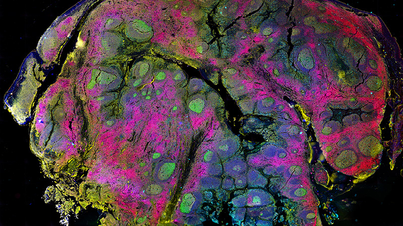
In this paper, Szulczewski et al. report a novel strategy to non-invasively characterize the cellular metabolism as well as to identify the macrophage population in the intact mammary tumor microenvironment of a mouse, by studying the endogenous fluorescence of metabolic co-factors NADH and FAD multiphoton and fluorescence life time imaging (FLIM).

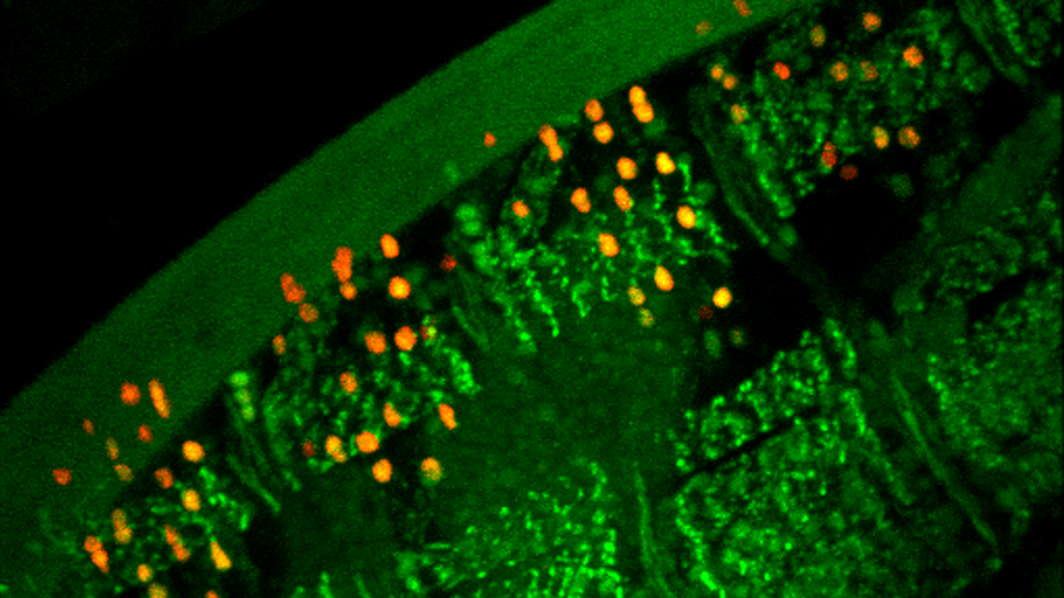
In particular, they could discriminate between tumor cells (with high autofluorescent NADH signal intensity) and stromal cells (with high endogenous FAD fluorescence intensity, predominantly phagocytic macrophages). Stromal collagen surrounding the tumor could also be imaged by second harmonic generation.

By taking advantage of Fluorescence Lifetime Imaging Microscopy, the cellular metabolism of the two different cell types has been characterized, showing that the FAD bright cells display a highly glycolytic NADH-FLIM signature, with a significantly shorter NADH average fluorescence lifetime with respect to the tumor cells. These results demonstrate that the glycolytic metabolic heterogeneity between monocytes and tumor cells can be used for imaging contrast.

In conclusion, this paper reports for the first time a novel way to use intravital, live, metabolic imaging in vivo to non-invasively and quantitatively identify distinct cell types within the breast tumor microenvironment, with high spatial and temporal resolution.

The relevance of this new label-free FLIM-based metabolite imaging approach in vivo is linked to the opportunity to be exploited as a readout for clinical diagnostics in the future, as several studies have demonstrated a correlation between NADH-FLIM changes and disease progression in biopsied colon, breast and melanoma tissue.