Scattering SNOM
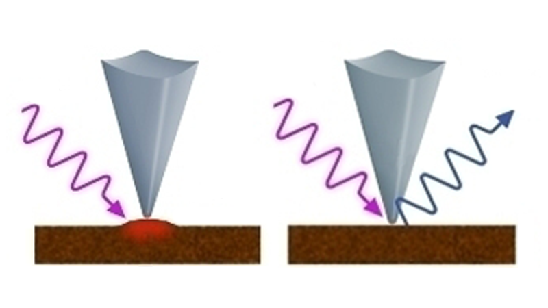
Infrared Scattering Scanning Nearfield Optical Microscopy (s-SNOM)
This technique provides information about the complex optical properties of the nanoscale region of the sample under a metallized tip. Specifically, both the optical amplitude and phase of the scattered light can be measured. With appropriate models, these measurements can estimate the complex optical constants (n, k) of the material. Additionally, the optical phase versus wavelength provides a good approximation to a conventional IR absorption spectrum usually grazing Incidence.
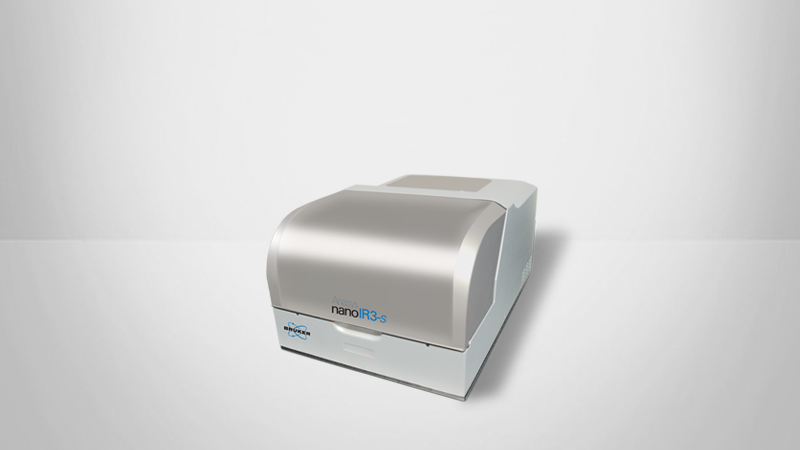
The s-SNOM technique works on a variety of materials, but the best signal to noise tends to be on harder materials with high reflectivity, high dielectric constants, and/or strong optical resonances. Bruker's nanoIR3-s provides an ideal platform for s-SNOM capabilities, eliminating the need for complex optical alignments:
- Patented adaptive beam steering and all reflective optics enables broad wavelength compatibility while eliminating realignment and refocusing at different wavelengths
- Patented dynamic power control maintains optimal power and signal over broad range of sources, wavelengths and samples
- Pre-mounted probes and motorized tip, sample and source alignment eliminates tedious steps in probe installation and re-optimization

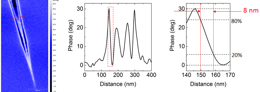
10nm Spatial Resolution Chemical Imaging and Spectroscopy
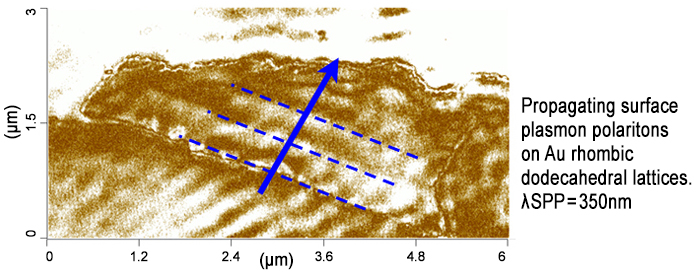
Graphene Plasmonics

High-Resolution Property Mapping
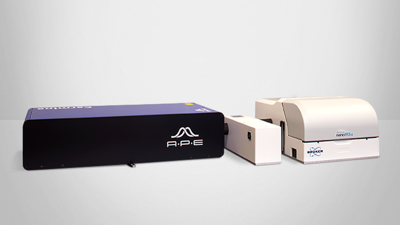
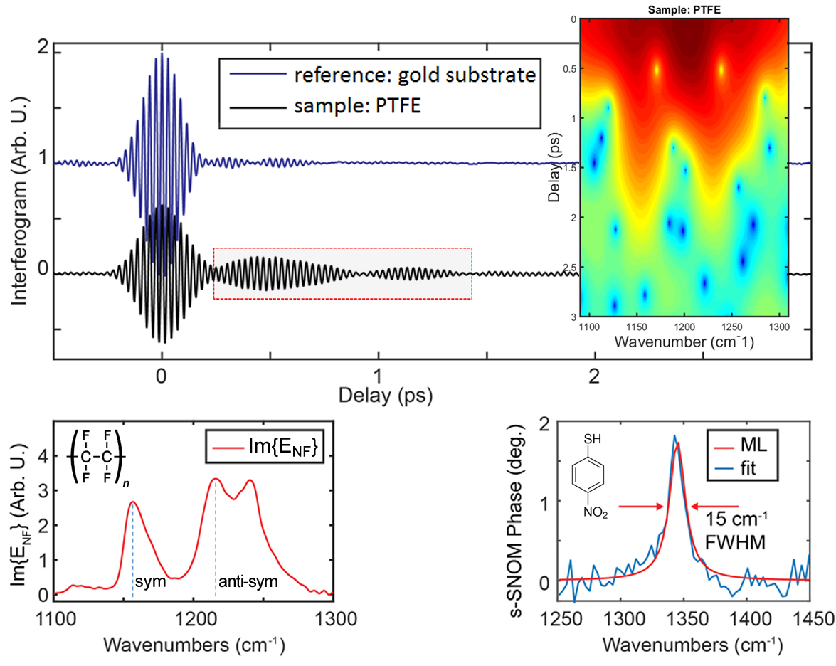
Highest Performance Nano FTIR Spectroscopy
Highest performance IR SNOM spectroscopy with the most advanced nanoIR laser source available.
- nano FTIR spectroscopy with integrated DFG, continuum based laser source
- Broadband synchrotron light source integration
- Multi-chip QCL laser source for spectroscopy and chemical imaging
Combine S-SNOM and AFM-IR to Create Remarkable New Data
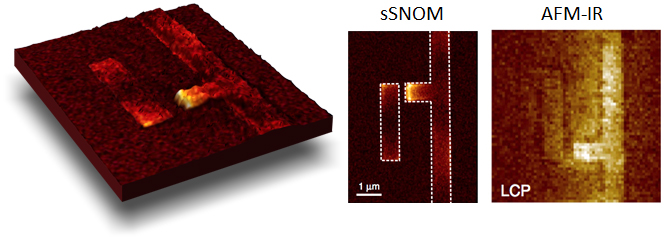
Complementary AFM-IR and Scattering SNOM images reveal, for the first time, the microscale origins of optical chirality on plasmonics structures. By accessing both the radiative (s-SNOM) and non-radiative (AFM-IR) information on plasmonics structures, unique and complementary plasmonic properties can be obtained.
nanoIR3-s Extends Beyond nanoIR to Visible and THz and Synchrotron Beam
- nanoIR3-s enables visible SNOM imaging
- System supports THz imaging and spectroscopy
- Special design available for use in synchrotron
- Easy change over of laser set up to maximize measurement time
- Simple swap out of optics components and detectors
Eliminating the Need for Complex Optical Alignments
- Patented adaptive beam steering and all reflective optics enables broad wavelength compatibility while eliminating realignment and refocusing at different wavelengths
- Patented dynamic power control maintains optimal power and signal over broad range of sources, wavelengths and samples
- Pre-mounted probes and motorized tip, sample and source alignment eliminates tedious steps in probe installation and re-optimization