Bioimaging with Light-Sheet Microscopy: Ultra Gentle Imaging of Living Samples
Ultra-gentle imaging of a multitude of live samples
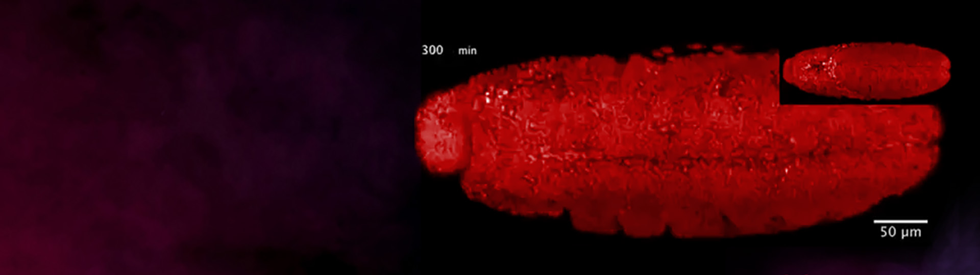
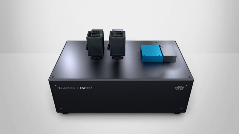
In this webinar, we discuss the benefits of light-sheet fluorescence microscopy as a gentle bio-imaging technique that is adaptable to a variety of samples. Specifically, we focus on the Luxendo InVi-SPIM with its unique light-sheet geometry designed for imaging live specimens in high resolution. We describe our innovative sample mounting technique and how we can use this to image cell lines, developing embryos, whole organisms, and more.
Webinar Summary
In this webinar, you can expect to learn the advantages of light-sheet fluorescence microscopy and how this technique can be adapted to image a multitude of different samples without the phototoxicity that plagues traditional imaging techniques such as spinning disk and confocal microscopy. Key topics discussed include:
Issues with Current Imaging Systems
- Balancing resolution and speed
- Photobleaching and phototoxicity during long time courses
- Imaging large volumes with sufficient time resolution
An Introduction to Light-sheet Microscopy:
- What is light-sheet fluorescence microscopy and how does it work?
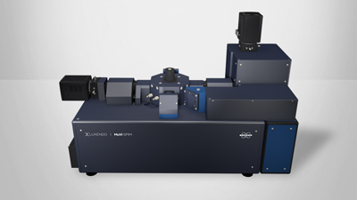
- Luxendo product introduction
Applications
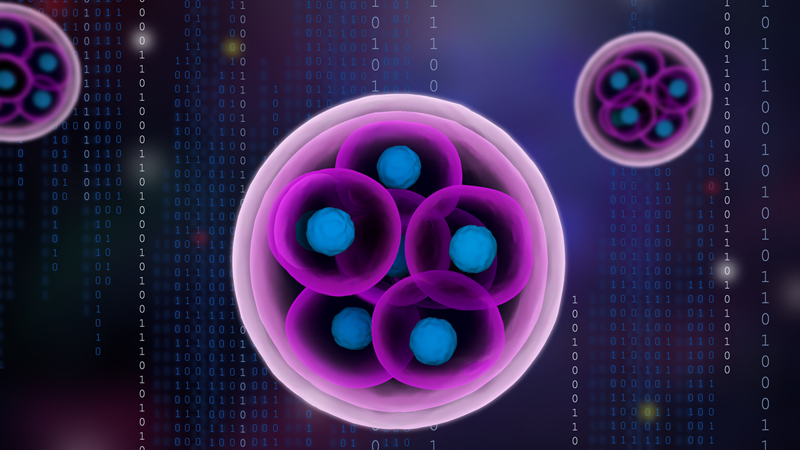
- Single particle tracking
- Embryo development
- IVF research
- Neuroscience
Find out more about the solutions featured in this webinar or our other solutions for Light-Sheet Microscopy:
Featured Products and Technology
Speaker

Dr. Dane Maxfield
Sales and Applications Specialist at Bruker Nano Surfaces
Dr. Dane Maxfield graduated with a B.A in Mathematics and Biology from Concordia University - St. Paul, before going on to complete a master’s degree in Applied Mathematics and a Ph.D. in Biology at the University of Utah.