Cleared Sample Imaging by Means of Light-Sheet Fluorescence Microscopy
Reveal new insights by imaging cleared samples with light-sheet microscopy
During this webinar, attendees learned about the advantages and applications of using light-sheet microscopy for imaging cleared samples. Key topics discussed include:
- Working components of light-sheet fluorescence microscopy
- Advantages of light-sheet microscopy over conventional fluorescence microscopy techniques
- Light-sheet microscopy for cleared-sample imaging
- Applications in neuroscience and developmental biology
Webinar Summary
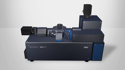
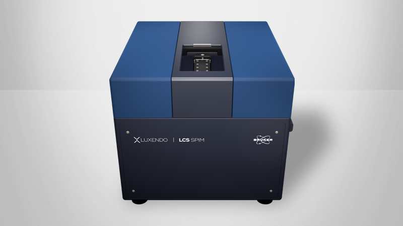
Light-sheet fluorescence microscopy serves to image a great variety of large biological samples. It works by having a sheet of laser light illuminate only a thin slice of a fluorescently labeled sample. Decoupling the illumination from the detection, typically via an orthogonal arrangement, enables fast, true volume, in-depth imaging with very low photodamage and bleaching effects.
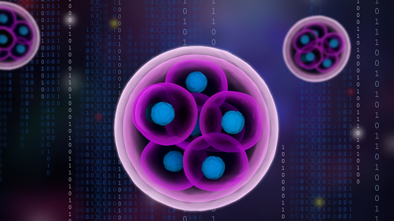
Clearing techniques have become a valuable tool for applications in 3D microstructure analysis of tissues. They modify the optical properties of usually opaque samples to render them transparent while keeping their structure and fluorescent labels intact. Clearing techniques can be combined with light-sheet microscopy to enable fast, long-term, confocal-like optical sectioning, and high-quality 3D imaging of cleared samples.
Find out more about the technology featured in this webinar or our other solutions for Cleared Sample Imaging:
Featured Products and Solutions
Speaker

Dr. Dane Maxfield
Sales and Applications Specialist at Bruker Nano Surfaces
Dr. Dane Maxfield graduated with a B.A in Mathematics and Biology from Concordia University - St. Paul, before going on to complete a master’s degree in Applied Mathematics and a Ph.D. in Biology at the University of Utah.