Palaeontology & Fossil Analysis with the M4 TORNADO
Why Use the M4 TORNADO for Fossil Analysis?
Fossils are the preserved record of life on Earth. The compositions of fossils may reveal much about the growth and environment of an organism while alive, and alteration that may have occurred after death during burial, mechanical reworking, and diagenesis.
Micro-XRF elemental mapping with the M4 TORNADO offers a non-invasive method for rapid compositional characterization of fossils of all kinds, providing more than just pretty pictures!

Alteration of Carbonate Fossil Materials During Diagenesis
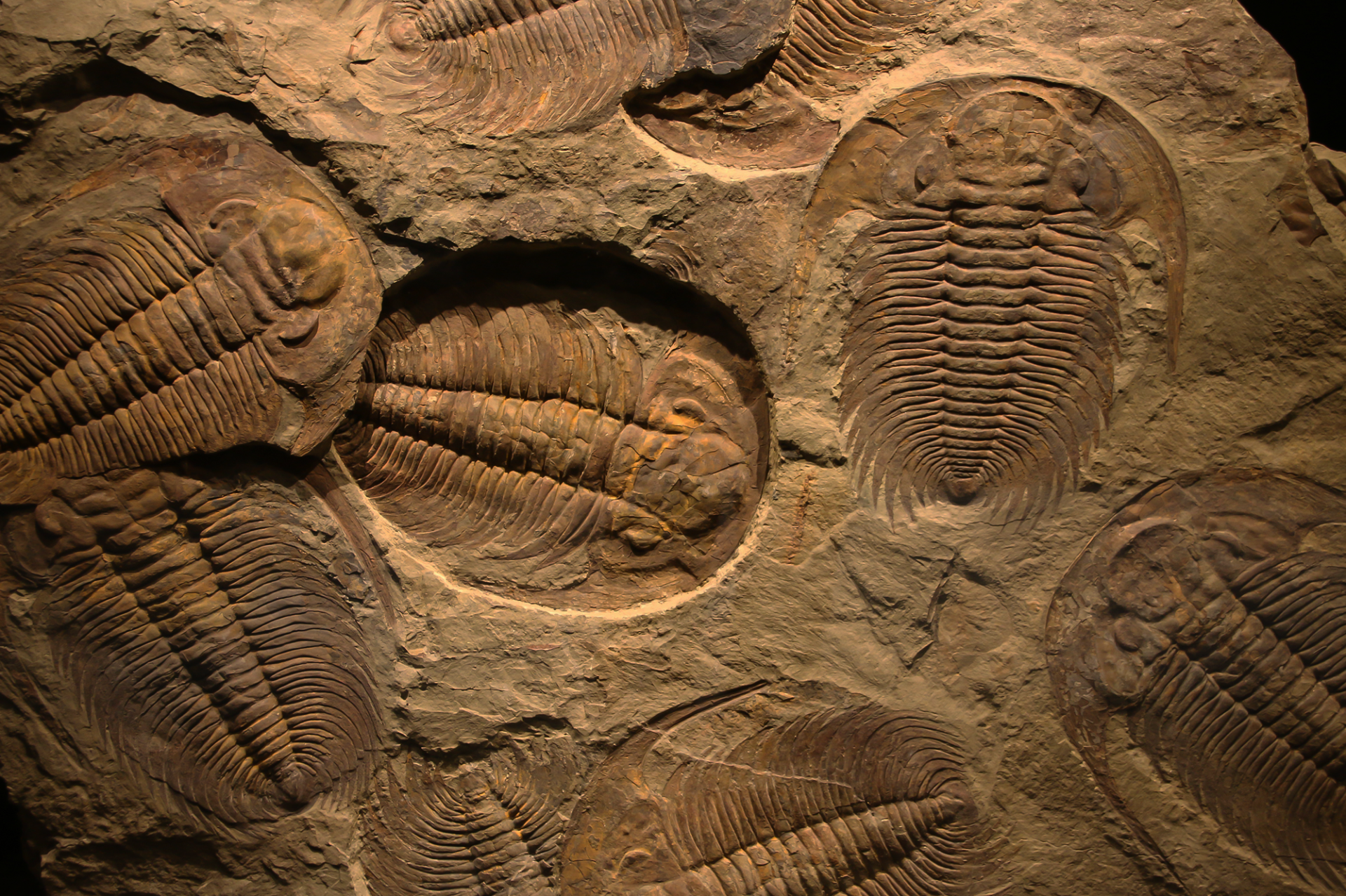
Fossils, while increasingly becoming collectors’ items, are our window into the evolution of life on Earth. Due to long residence times buried in sediments, the primary minerals are altered and in the case of organic materials usually completely lost.
With replacement of the primary structures, chemical fingerprints may provide a means to image the structures of fossils, and understand the alteration processes.

Geochemical Fingerprints of Fish Fossils
At several locations around the globe spectacularly preserved Eocene fish fossils are found that provide a window into the climatic and geological history of this epoch of Earth history. Although well preserved, visual imaging (by camera or microscope) may require regular wetting of the fossil (e.g., with alcohol). An alternative method is through non-invasive micro-XRF elemental mapping.
Elemental maps, such as those collected by the M4 TORNADO micro-XRF spectrometer, provide key information such as a detailed characterization of anatomical structures, which may otherwise be problematic or impossible to discern, but important for taxonomic, phylogenetic and palaeo ecological studies. In addition, the deeper penetration of X-ray allows imaging of some features hidden by overlying structures or thin layers of sediment. The rapid data collection provided by the M4 TORNADO also allows collections from different locations to be measured and systematically compared with limited need for additional handling of the fossils.

Elemental Characterization of Vertebrate Fossils
Elemental mapping by micro-XRF allows a palaeontologist to dig deeper into the anatomy of vertebrate fossils, revealing information about differences in the compositions of different bones, including where burial and time have caused alteration to the original bone compositions and therefore which areas of the fossil represent original growth histories.
The non-invasive measurement approach afforded by the M4 TORNADO micro-XRF spectrometer allows such detail to be revealed without damaging the precious fossils specimens and providing a data set that can be accessed remotely by other researchers that may not have in-person access.
The <20 um X-ray spot and high sensitivity detectors allows intricate detail to be visualized, e.g., in the bat‘s jawbone, the shoulder and the feet with part of the tail (lower middle maps).

Deformation Feature or Ancient Fossils? Mapping Elemental Variations in Stromatolite Structures
Stromatolites, layered sedimentary structures that formed by microbial mats trapping and binding sediment, represent the earliest preserved physical evidence of life on Earth. Therefore, significant controversy exists around the earliest occurrence of stromatolites, with several publications arguing for and against structures preserved in the Isua Supercrustal Belt of Greenland, that could be evidence for life as far back as c. 3.81 billion years ago.
A key piece of evidence required for positive identification of stromatolites is a layered structure (e.g., the stromatolite maps to the right). Micro-XRF provides a means for seeing through potential alteration of this original structure during long crustal residences, which may no longer be visible to the naked eye, through micro-scale characterization down to trace element levels. When correlated with other evidence (e.g., mineralogy, isotopes) we may build a window into the deep past and make robust interpretations about the early evolution of the Earth.
Learn more about the study here:

See the Data and Images in More Detail


Interested in Transforming Your Lab with the Power of the M4 TORNADO?
Whether you're exploring new instrumentation, planning your next grant proposal, or simply curious about how micro-XRF could fit into your workflow - we're here to help.
Explore How the M4 TORNADO Supports Your Research - Let's talk
Connect with our regional team to discuss your geoscience applications. We’ll help align your goals and route next steps to the right Bruker specialists. Book a short, no‑obligation meeting to get started.
Request Pricing
Get a customized quote based on your lab’s needs and configuration preferences.
Request a Live Demo
See the M4 TORNADO in action with real academic use cases and sample workflows.