High-field, high impact: How NMR collaboration at CERM, University of Florence is enhancing Covid-19 protein research
Within an international collaboration of over 50 NMR experts, a team led by Prof. Dr. Lucia Banci at CERM, University of Florence, Italy, has been helping to determine the structures of proteins found in SARS-CoV-2.
Thanks to the recent installation of Bruker’s 1.2 GHz nuclear magnetic resonance (NMR) spectrometer, she has been able to provide detailed information about atomic-level interactions, and so contribute to the development of innovative approaches for drug design and optimization, for antibody validation, including those against SARS-CoV-2, and for high order structure (HOS) of biological drugs.
The academic story of Prof. Dr. Lucia Banci is one of evolution on two fronts – evolution of the capabilities of NMR instruments she has used, and a parallel evolution in the scale of the molecular systems studied. As a case in point, the 1.2 GHz system she is using today bears little resemblance to the 60 MHz instrument she first used in the 1970s, while her recent work on the structure and interactions of 30-40 kDa proteins would have been unimaginable in the days when the technology was limited to small, simple molecules and complexes.
The essential role of metal ions in biochemistry
A single topic unites much of Prof. Banci’s research over the last 45 years – metal ions. These, she strongly believes, must be understood in depth to achieve progress in many areas of Life Sciences. As she points out, “More than one-third of proteins require metal ions to function and are at the heart of many biological processes. Trafficking of metal ions is tightly controlled and often mediated by weak, transient protein–protein interactions.” This conviction is borne out of her long-standing interest in the field – from being the first to determine the solution structure of a paramagnetic metalloprotein1 (at the time thought to be impossible), to devising molecular dynamics approaches for metal-containing proteins2 and uncovering the details of copper transfer in cytochrome C oxidase3.
Underpinning all this is Prof. Banci’s background in inorganic chemistry, which initially involved using electron paramagnetic resonance (EPR) to investigate the magnetic properties of metal ions and their complexes. It wasn’t long, however, before she discovered the power and versatility of NMR: “In the early days of research, if you wanted to determine structures with atomic-level resolution, the conventional method was X-ray crystallography. But that required crystals, whereas NMR seemed much more versatile, with emerging experimental techniques opening up the possibility of determining complex structures in solution.”
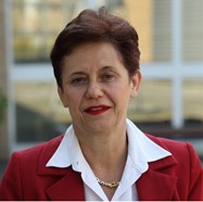
Prof. Dr. Lucia Banci is Professor of Chemistry at the University of Florence, and one of the founders and Director of the Center of Magnetic Resonance (CERM). She is also the Head of the Italian Core Center of the Integrated Structural Biology Infrastructure’s Research Infrastructure Consortium (Instruct-ERIC), and a member of the Instruct-ERIC Executive Committee and Council.
The versatility of NMR for structural biology research
That early realization proved to be spot on, foretelling as it did the growth in experimental protocols for NMR, and the detailed insights that they enable today. From the simple small-molecule studies that were the cornerstone of Prof. Banci’s early research she has seen – and in many cases contributed – to many of the most exciting applications since. These include the use of NMR to characterize drugs, metalloproteins, vaccines, new materials, RNA, and solid-state materials, and to understand how large complexes interact, and how proteins fold, mature, and take up metals.
“Even just within the biological field, NMR has an enormous breadth of application”, she says. “We can study systems that are floppy, mobile, interacting, and – something that was a particular breakthrough for me – molecules in living cells”4. The latter approach allows individual proteins or protein complexes to be studied in living human cells, and Prof. Banci was instrumental in developing this field, specifically for the efficient expression and labeling of proteins directly in human cells, thus characterizing human proteins in the human cells where they have been produced. This approach allowed the description and the rationalization of several processes, from metal ion uptake and its impairment, as it happens in pathological conditions, to factors determining the redox states of proteins and the processes of protein folding.
As Prof. Banci says: “In-cell NMR bridges the gap between two areas. Biological studies maintain the cellular environment but lack atomic-level information, whereas conventional structural characterization provides detail about the molecular structure but lacks the biological context. What’s special about in-cell NMR is that it allows us to obtain detailed structural information about a biomolecule when it is in its natural environment. This can be very helpful in the early stages of drug development, and for screening in particular5. For example, we can see how proteins bind a drug, or examine why a protein may bind one drug preferentially over another.”
Excitement and challenges: Installing the new Bruker 1.2 GHz instrument
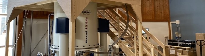
In parallel to this NMR application development is the evolution in instrument power, and Prof. Banci enthusiastically describes the Bruker 1.2 GHz instrument that was installed at the Center of Magnetic Resonance (CERM) in 2020, bringing the total number of instruments at the facility to 12.
“Getting our new instrument was really exciting – it was the first installation of this instrument globally and was very much in the spotlight. It was also quite challenging, as shortly after installation began, the pandemic lockdown restrictions came into force. We had to deal with logistical problems such as reduced staff numbers, delayed helium deliveries and even disruptions to catering arrangements – but with the expert assistance of the Bruker engineers we succeeded, and by the start of April the instrument was ready to go.”
Prof. Banci explains that as well as being used for her own research into Covid-19 protein structures and antibodies, the 1.2 GHz instrument is available as a service to other groups in the EU. “We started acquiring spectra on our new system straight away – so even while lockdown continued, we were receiving samples, acquiring data, and sending results. The instrument hasn’t stopped – and I don’t think we have either!”
Through it all, she has been very happy with the performance of the system: “The spectra are beautiful,” she says. “The high magnetic field, combined with the cryoprobe, provides exquisite resolution and sensitivity. This means that we can work at concentrations approaching those found in living cells, and so obtain insights that are more biologically relevant.”
Fast and efficient collaboration on Covid-19
A major application of the 1.2 GHz instrument at CERM is to discover more about the structures of the proteins contained in the SARS-CoV-2 virus, as part of the Covid-19 NMR Project. This is a consortium of over 50 research groups from across the world that aims to characterize the RNA and proteins of SARS-CoV-2 using NMR spectroscopy, and in doing so assist the development of drugs to treat Covid-19.
The consortium is led by Prof. Dr. Harald Schwalbe at Goethe-Universität in Frankfurt, Germany, who Prof. Banci had known for many years previously: “When the pandemic started, I immediately realized the potential for NMR to address several research areas associated with the virus. I know Harald well, and after discussions we agreed that there was much to be gained by setting up a consortium to tackle these questions using NMR.”
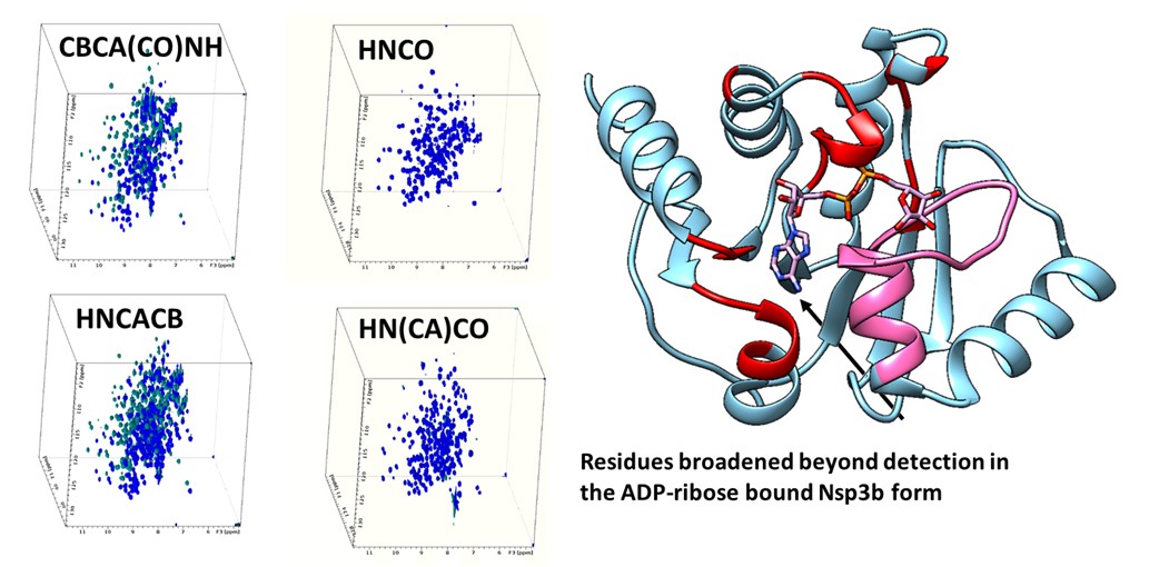
She explains that through the consortium, they have been able to set up effective protocols for the various steps of protein and RNA structure determination – including production, labeling, NMR data acquisition and assignment. She says: “Thanks to this, we now have a process for addressing questions around new variants in as little as two weeks, which is a major outcome for us. In addition, using the 1.2 GHz Bruker system, we are now characterizing SARS-CoV-2 proteins in detail6,7, and examining the effect of both natural ligands and existing drugs, to see if they block progression of the virus.”
Carrying out this work within the consortium has made progress much faster than it would otherwise have been, she explains: “By exchanging materials, experimental protocols and results, and by working out how to put our expertise and instruments to best use, we can work synergistically. It’s a very effective and time-efficient way of getting results.”
Crucial to much of this success, says Prof. Banci, is data management. “To respond to urgent research questions like those raised by Covid-19, we need to react quickly, and that means making data available to other researchers rapidly. But at the same time, it must be managed and archived correctly – the right data is of no use if it is not organized and retrievable. This is especially relevant to NMR because of the very large datasets generated from studies of biomolecules – and I think it is an aspect that the scientific community generally needs to consider carefully. To advance knowledge quickly, we need efficient systems for managing scientific data and sharing it with those who need it.”
Future research: virus–host interactions and new SARS-CoV-2 variants
Looking back over the last 18 months, Prof. Banci concludes that the consortium continues to be a very successful example of how a broad network of enthusiastic scientists can collaborate to help understand a specific problem. “I think that the example of Covid-19 NMR should be followed elsewhere,” she says. “By combining our strengths, we can achieve much more than would be possible within an individual research group.”
And the work continues, with further insights into SARS-CoV-2 in the pipeline: “The next step is to go deeper into the interactions between the components of the virus and its host, and to address questions around variants and mutants.”
She concludes: “We’ve shown through this consortium that NMR can address multi-faceted problems, by providing the atomic-level resolution needed to really understand what’s going on at a molecular level. The versatility of NMR is its key strength, and I’m confident that this remarkable technique will continue to provide major scientific insights long into the future, across many fields of study.”
For more information about the work of the Banci group, please visit https://www.cerm.unifi.it/about-us/people/lucia-banci
For more information about the Covid-19 NMR Consortium, please visit https://covid19-nmr.de/
References
- Banci L et al. The three-dimensional structure in solution of the paramagnetic protein high-potential iron-sulfur protein I from Ectothiorhodospira halophila through nuclear magnetic resonance, Eur J Biochem, 1994; 225: 715-725.
- Banci L, et al. Mitochondrial copper(I) transfer from Cox17 to Sco1 is coupled to electron transfer, Proc Natl Acad Sci USA, 2008; 105: 6803-6808.
- Banci L, et al. Molecular dynamics of metallo proteins, In: Molecular Modelling and Dynamics of Bioinorganic Systems, Dordrecht, The Netherlands: Kluwer academic publishers, 1997.
- Banci L, et al. Atomic-resolution monitoring of protein maturation in live human cells by NMR, Nat Chem Biol, 2013; 9: 297-299.
- Luchinat E, et al. Drug screening in human cells by NMR allows early assessment of drug potency, Angew. Chem. Int. Ed., 2020; 59, 6535 –6539.
- Cantini F, et al. 1H, 13C, and 15N backbone chemical shift assignments of the apo and the ADP-ribose bound forms of the macrodomain of SARS-CoV-2 non-structural protein 3b. Biomol NMR Assign, 2020; 14: 339–346.
- Gallo A, et al. 1H, 13C and 15N chemical shift assignments of the SUD domains of SARS-CoV-2 non-structural protein 3c: “The SUD-M and SUD-C domains”, Biomolecular NMR Assignments, 2021; 15: 165–171.
About CERM
The Center of Magnetic Resonance (CERM) is a center for research, knowledge transfer, and higher education of the University of Florence, located at the Polo Scientifico (Scientific Campus) in Sesto Fiorentino. The Center is a research infrastructure for NMR in the Life Sciences supported by the European Commission, by the Italian Ministry for University and Research, and by the Regional Tuscany government. . CERM is the Italian center of the ESFRI Landmark Research Infrastructure Instruct-ERIC. CERM shares its infrastructure with the Interuniversity Consortium Magnetic Resonance of Metal Proteins (CIRMMP), covering an area of 3,000 square meters and hosting a number of laboratories, offices, and common rooms. The flagship of the Center is the impressive collection of NMR spectrometers which feature the largest magnetic field range in the world (1.2 GHz) and ranks it among the best equipped laboratories in the world.
For more information about CERM, please visit https://www.cerm.unifi.it/.
About Bruker Corporation
Bruker is enabling scientists to make breakthrough discoveries and develop new applications that improve the quality of human life. Bruker’s high-performance scientific instruments and high-value analytical and diagnostic solutions enable scientists to explore life and materials at molecular, cellular, and microscopic levels. In close cooperation with our customers, Bruker is enabling innovation, improved productivity, and customer success in life science molecular research, in applied and pharma applications, in microscopy and nanoanalysis, and in industrial applications, as well as in cell biology, preclinical imaging, clinical phenomics and proteomics research and clinical microbiology.
For more information, please visit: www.bruker.com.