
Inhibitory Control of Dopaminergic Neurons
Multiphoton imaging enables insights into neuronal behavior
Attendees will learn about Dr. Rebekah Evan’s work with multiphoton imaging to explore the inhibitory control of dopaminergic neurons and their impact on neurodegenerative conditions such as Parkinson’s Disease. She will explain her use of various techniques such as:
- Two-photon calcium imaging;
- Optogenetic activation;
- Whole-cell patch clamp analysis; and
- Computational modeling.
Presenter's Abstract
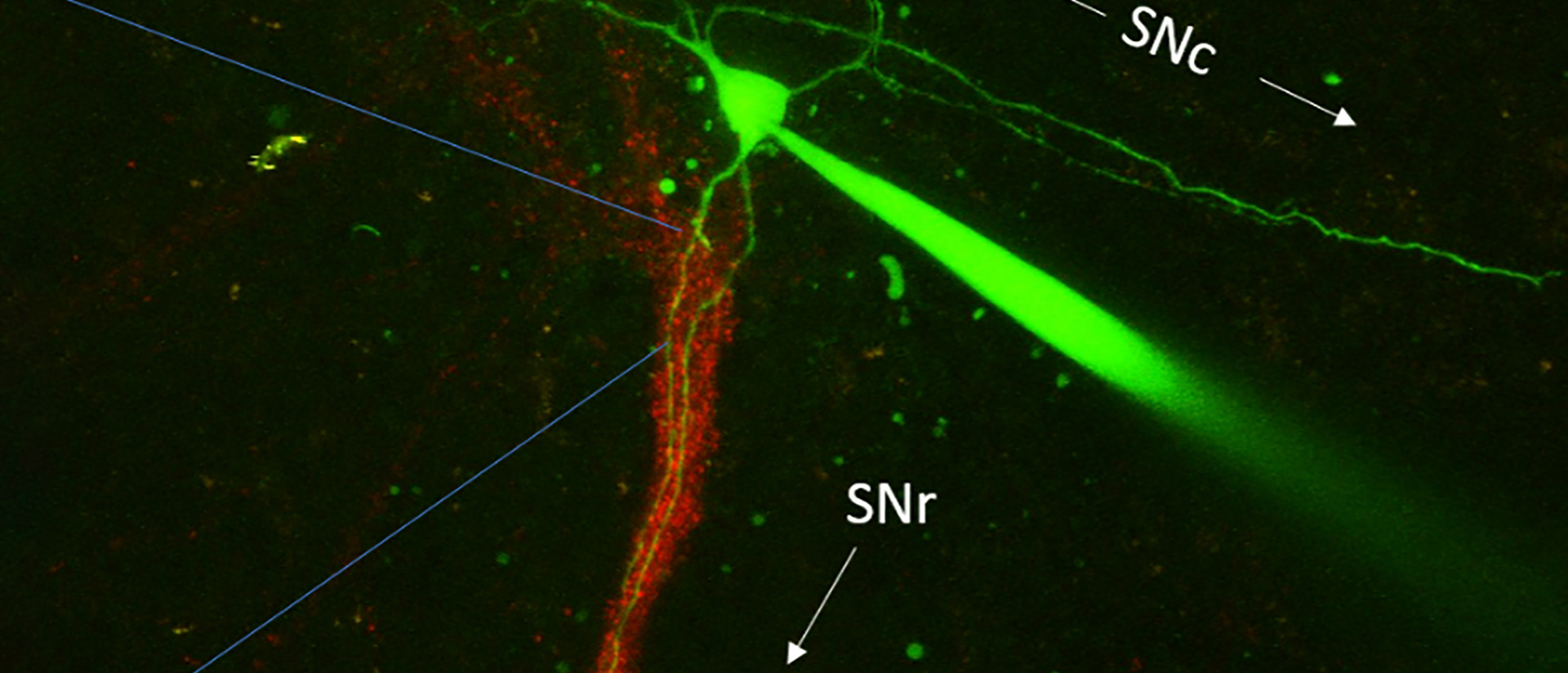
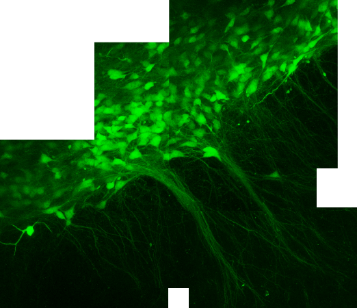
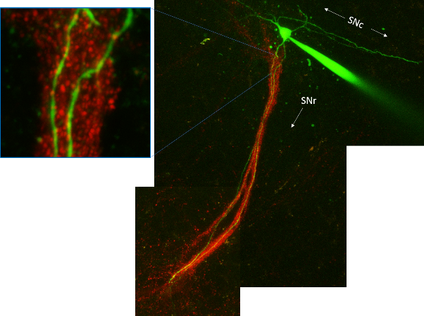
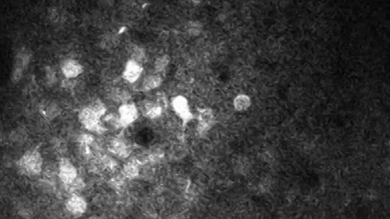
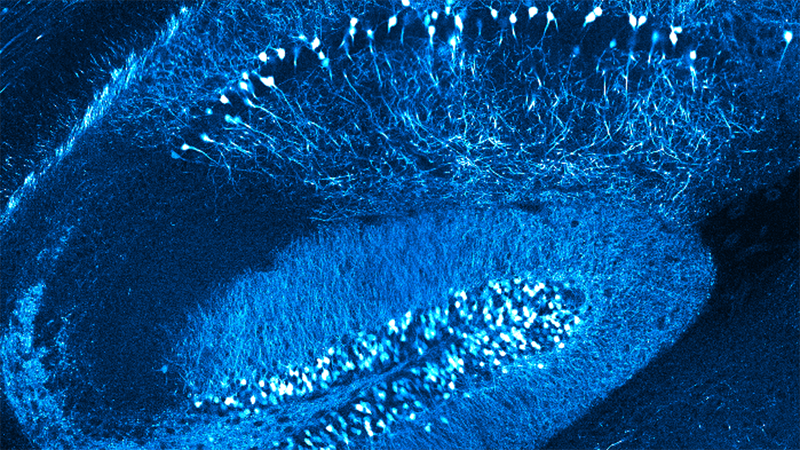
Dopamine neurons are embedded in a predominately inhibitory network and because dopamine neurons are tonically active, both excitatory bursts and inhibitory pauses in their firing can transmit meaningful information. In this talk, I will address how these bursts and pauses in activity interact with each other through the recruitment of specific intrinsic and synaptic currents. I will show that two populations of substantia nigra dopamine neurons, defined by their differential vulnerability to neurodegeneration in Parkinson’s Disease, differ in their dendritic calcium signaling and their ability to recover from periods of inhibition. I use dendrite-specific optogenetic activation and two-photon calcium imaging to functionally map the location of inhibitory synaptic inputs onto dopamine neuron dendrites. I also use whole-cell patch clamp and computational modeling to show how distinct inhibitory sources differentially modulate dopamine neuron activity. Overall, I describe a striking heterogeneity in the cellular and circuit level control of dopaminergic signals.
See More of This Presenter's Research
- Evans RC, Zhu M, Khaliq ZM. Dopamine Inhibition Differentially Controls Excitability of Substantia Nigra Dopamine Neuron Subpopulations through T-Type Calcium Channels. J Neurosci. 2017 Mar 29;37(13):3704-3720. doi: 10.1523/JNEUROSCI.0117-17.2017.
- Evans RC, Twedell EL, Zhu M, Ascencio J, Zhang R, Khaliq ZM. Functional Dissection of Basal Ganglia Inhibitory Inputs onto Substantia Nigra Dopaminergic Neurons. Cell Rep. 2020 Sep 15;32(11):108156. doi: 10.1016/j.celrep.2020.108156.
Featured Products & Technology
Speaker

Rebekah Evans Ph.D., Georgetown University
Dr. Rebekah Evans is an Assistant Professor in the Department of Neuroscience at Georgetown University, Washington D.C. She received her Ph.D. degree in neuroscience from George Mason University under Dr. Kim Blackwell studying calcium dynamics and signaling pathways underlying synaptic plasticity where she applied computational approaches. Her graduate studies were funded by an NRSA F31 from NINDS. Dr. Evans did her post-doctoral work at the National Institute of Neurological Disorders and Stroke with Dr. Zayd Khaliq. Dr. Evans recently received a K99/R00 award from the BRAIN Initiative to functionally dissect inhibitory circuitry in the basal ganglia. She is an expert in two-photon calcium imaging, electrophysiology, and optogenetics and applies computational approaches to investigate the brain’s cells and circuitry most vulnerable in Parkinson's Disease.