Protein Allostery and NMR
Allostery – the phenomenon by which an event in one part of a molecule causes an effect in another – is a key feature of protein regulation in all living cells. Rather than directly affecting a protein’s active site, allostery can modulate the active state of a protein through ligand binding, covalent modification or mutation elsewhere in the protein.
This can change the tendency of the protein to bind certain ligands or alter its catalytic effects, and result in a shift in the equilibrium of protein conformations within a population. In biological processes allostery is involved in signal transduction, metabolism, enzyme activation and gene regulation.
However, despite the ubiquity of the phenomenon in biology and a 50-year quest to uncover how it works, still relatively little is known of the mechanisms by which allostery actually functions.
Communication networks
The current model of allosteric signaling within protein structures involves amino acid residue networks that are able to communicate with each other over long molecular distances, analogous to a telecommunications network. Early research was able to identify allosteric sites that were physically linked with active or second sites, and could induce conformational changes in the protein. But given the long distance over which these signals are able to communicate, it is clear that dynamically coupled residues play as much of a role in allosteric processes as physically coupled residues. Indeed, many allosteric interactions occur with imperceptible or no structural change at all.
Recent research using high-throughput experimental and computational methods has been able to identify allosteric binding sites and residue networks, but there are challenges. For example, these require a detailed prior structural and functional knowledge of the protein. Furthermore, the dynamic processes involved in allostery cover a very large timescale ranging from picoseconds to milliseconds, which can be difficult or impossible to capture with existing methods. And even a small shift away from the protein’s native state within a population can have a large impact on function, but these subtle changes may not be detectable by methods such as X-ray.
Over the last decade, nuclear magnetic resonance (NMR) spectroscopy has emerged as a technique that can overcome many of these obstacles. As well as being able to provide structural information on proteins in solution, NMR can also reflect dynamic processes across a broad timescale, and in sparse populations of conformational states. Thus it can detect allostery that would be difficult or impossible to identify using other methods.
Drawing the map
In a study in Scientific Reports, researchers led by Peter Wright at the Scripps Research Institute, La Jolla, California, demonstrated a method that uses NMR to identify allosteric communities and characterize the information flux between them.
The team used a eukaryotic kinase, p38γ MAPK, to carry out their experiments. The allosteric sites of this family of enzymes have been well characterized, but it is unknown how the regulatory sites communicate with catalytic residues. To explore this, the researchers mutated methyl-containing side chains within the protein at 20 specific sites. The effects of these mutations perturbs the allosteric network within the protein, which can be measured via changes in 1H-13C methyl chemical shifts.
Using triple-resonance NMR and network analysis approach, the team were able to develop a map showing communication paths between regulatory elements and catalytic sites. Because the method allowed them not just to look at the overlap between the different elements of the network, but the information flux between them, the team could demonstrate the functional importance of the various pathways. In this way, they identified critical nodes, with both a high level of connections and the greatest level of information flowing between them, which corresponded to known allosteric sites in the 3D structure, despite no structural information being needed as input.
The researchers say that the method will be valuable in future experiments, including for drug discovery, by delivering an information-rich and direct view of allosteric networks within proteins.
An advanced system
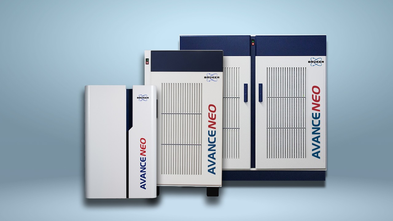
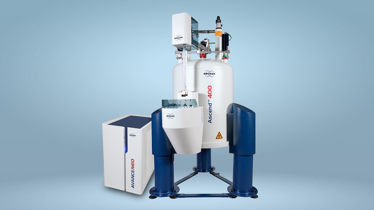
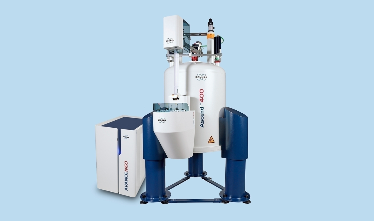
In their experiments, the researchers used Bruker Avance 750 and 900 MHz spectrometers equipped with triple-resonance gradient probes.
The Bruker Avance III HD offers fast, flexible NMR with the most advanced RF generation in the industry. And, Bruker’s Triple Resonance Probes are extensively used in experiments to determine the structure of macromolecules with NMR, delivering both performance and flexibility, including access to advanced NMR techniques.
References
- Aoto PC, Martin BT & Wright PE. NMR Characterization of Information Flow and Allosteric Communities in the MAP Kinase p38γ. Scientific Reports 2016; 6: 28655.
- Goodey NM & Benkovic SJ. Allosteric regulation and catalysis emerge via a common route. Nature Chemical Biology 2008; 4: 474-482.
- Kern D & Zuiderweg ERP. The role of dynamics in allosteric regulation. Current Opinion in Structural Biology 2003; 13: 748-757.
- Tsai C-J & Nussinov R. A Unified View of “How Allostery Works”. PLoS Computational Biology 2014; 10: e1003394.
- Tzeng S-R & Kalodimos CG. Protein dynamics and allostery: an NMR view. Current Opinion in Structural Biology 2011; 21: 62-67.