Live Cell Imaging
Live cell single-molecule localization microscopy using the Vutara
Biological imaging requires viewing dynamic structures within cells; either visualizing individual biomolecules in cells (particle tracking) or observing biological processes, such as membrane, organelle or cytoskeletal movement.
The Vutara VXL microscope's capabilities are ideal for live-cell single-molecule localization microscopy:
- 3D localization using patented biplane technology – each image has 1 µm of 3D data - without taking a Z series
- Axial range can be extended with a Z series
- Fast acquisition – up to 800 fps full frame, and up to 3000 fps with a smaller ROI
- Sensitive detector – cooled scientific CMOS camera
Particle Tracking
The Vutara VXL microscope is designed for single-molecule localization microscopy and is thus ideally designed for particle tracking experiments. Vutara’s intrinsic ability to image single fluorophores at high speed and in three dimensions makes it ideal for particle tracking. Samples can be labeled with organic dyes, proteins or quantum dots and imaged using the Vutara VXL's high-speed and sensitive camera. The Vutara's SRX software has built-in particle tracking algorithms for tracking fluorophores over multiple frames. This is a powerful and unique feature of single-molecule localization microscopy over image based super-resolution microscopes such as STED or SIM. The SRX software also includes a comprehensive particle tracking statistics package for analyzing your data.
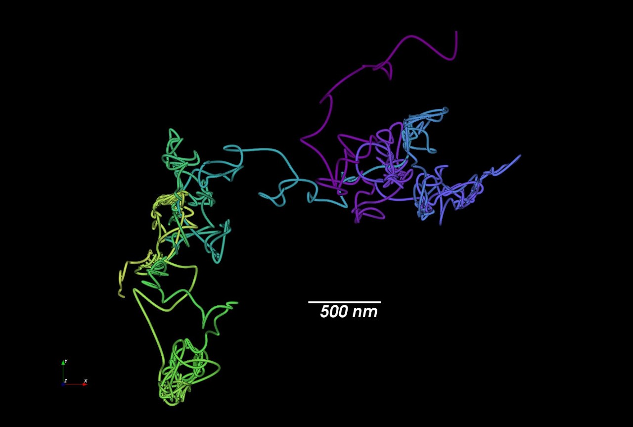
On the left we can see diffusion of a single EGF receptor labeled with a quantum dot.
Cellular Imaging
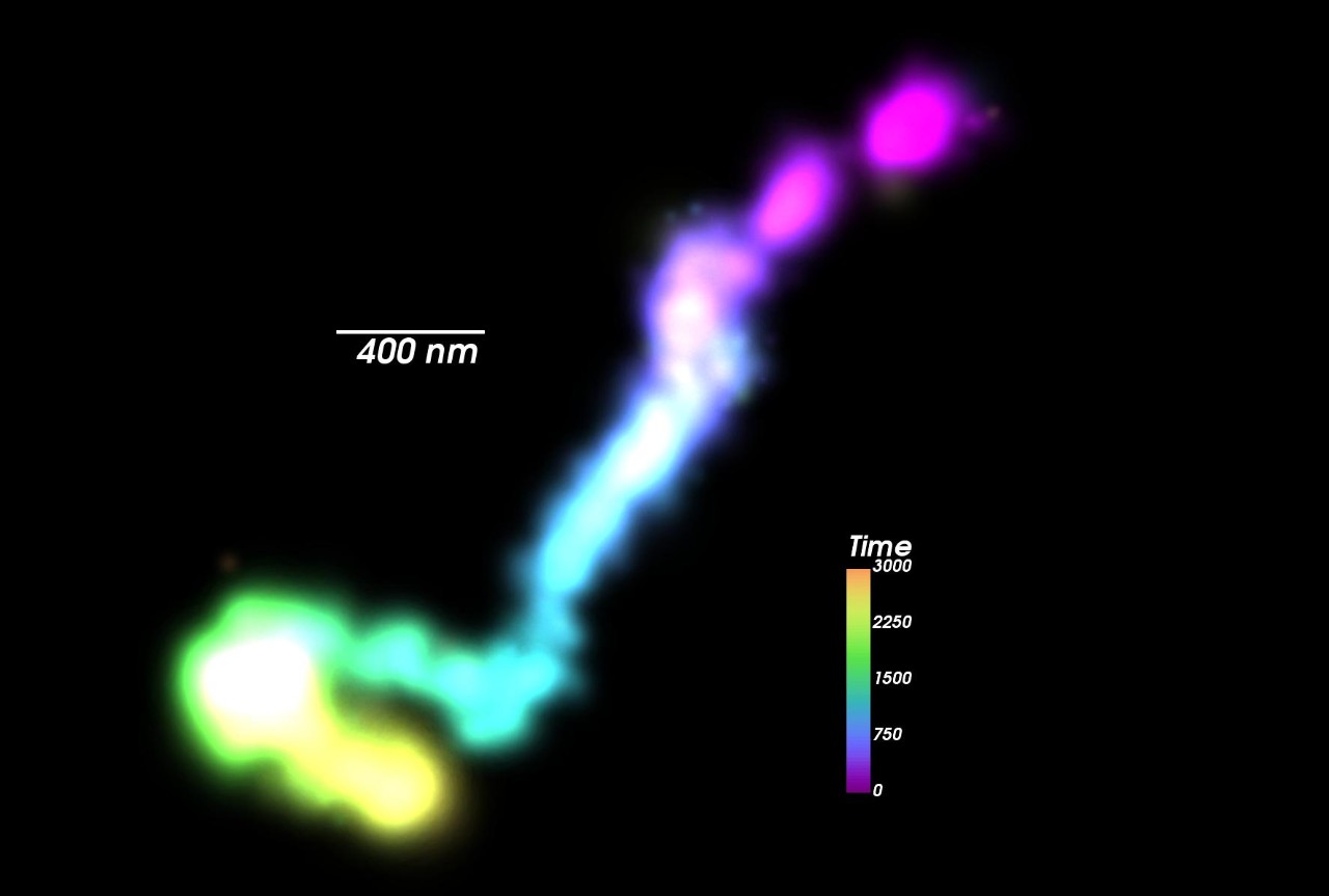
The Vutara VXL microscope is designed for single-molecule localization microscopy and is capable of capturing biological motion on a meaningful timescale. This unique imaging modality allows the user to collect dynamic super-resolution images of the structure of interest, while also giving the user the precise 3D position of each molecule over time. This highlights the unique imaging modality of single-molecule localization microscopy and sets its apart from imaged based super-resolution techniques.
On the right we see two experiments monitoring mitochondrial dynamics in live cells. One imaged with an orange HaloTag® dye (549) and the other labeled with a photactivatable far-red dye (PA-JF-646®).
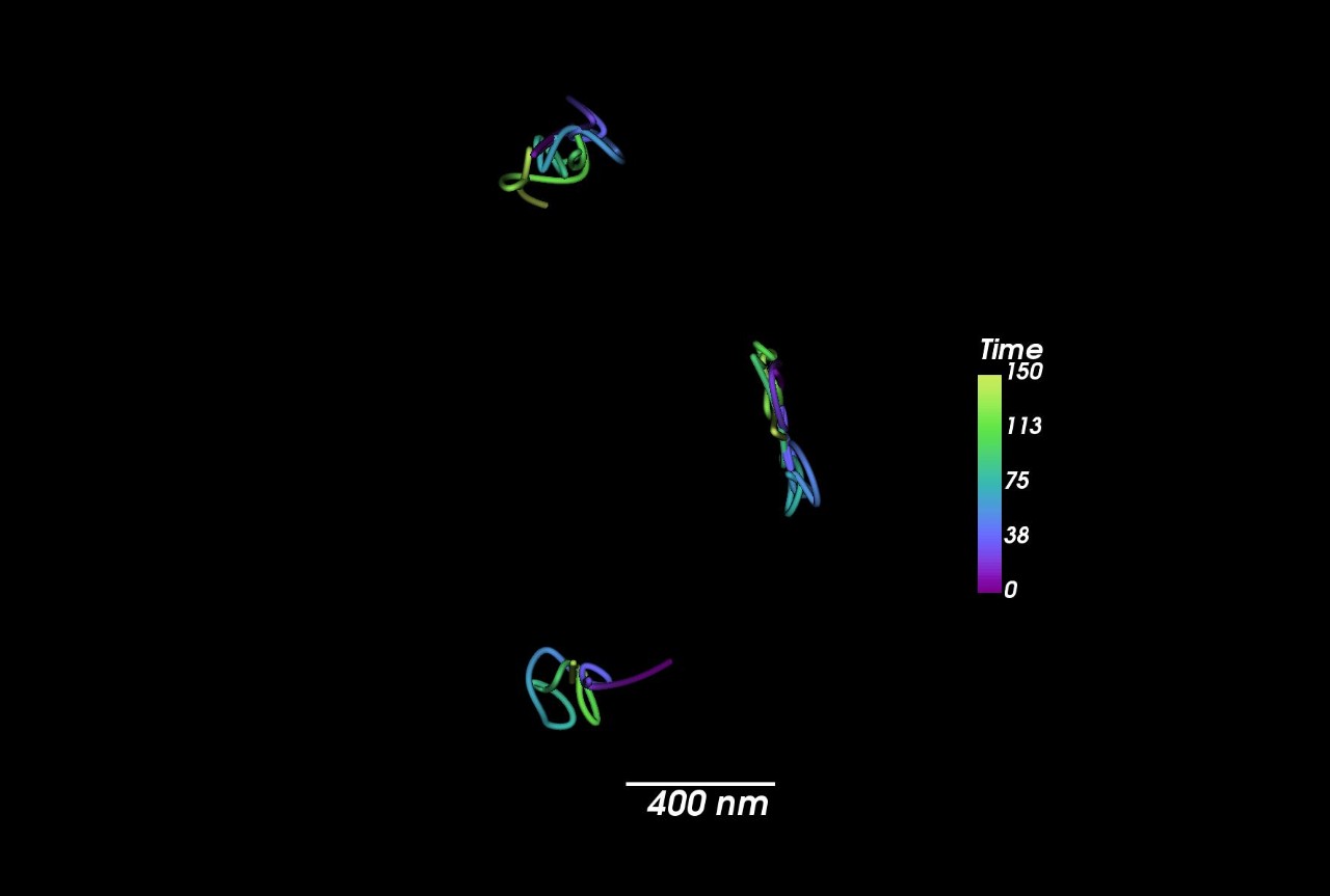
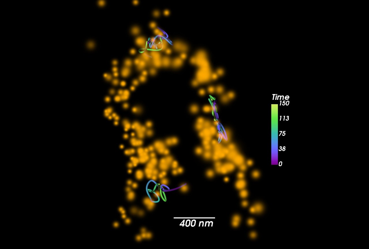
Combined Particle Tracking and Cellular Imaging
The Vutara VXL microscope's ability to perform multicolor single-molecule imaging allows for some unique experimental design. On the left is shown a two color experiment in cells expressing tomm-20::HaloTag®. In this experiment the tomm20::HaloTag® is labeled with a dense concentration of JF549® and a very sparse concentration of PA-JF646®. This allows the dense dye to be used as a contextual marker allowing the reconstruction and localization of mitochondria over time. While the second dye allows the tracking of the diffusion of single molecules in the context of the mitochondria. Using this technique one can monitor how the diffusion of proteins in a membrane changes as mitochondria traffic and undergo fusion or fission.