Unlocking Plant Biology with Advanced AFM
Learn about advanced, innovative uses of BioAFM technology.
In this webinar, guest experts Dr. Simoné Bovio (Ecole Normale Supérieure) and Prof. Gleb E. Yakubov (University of Leeds) discuss research into plant biology using bioAFM instrumentation.
- Dr. Bovio, from the Plant Reproduction and Development Laboratory at ENS, will speak on using AFM to assess turgor pressure in plants at the single-cell level.
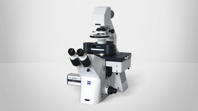
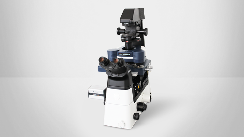
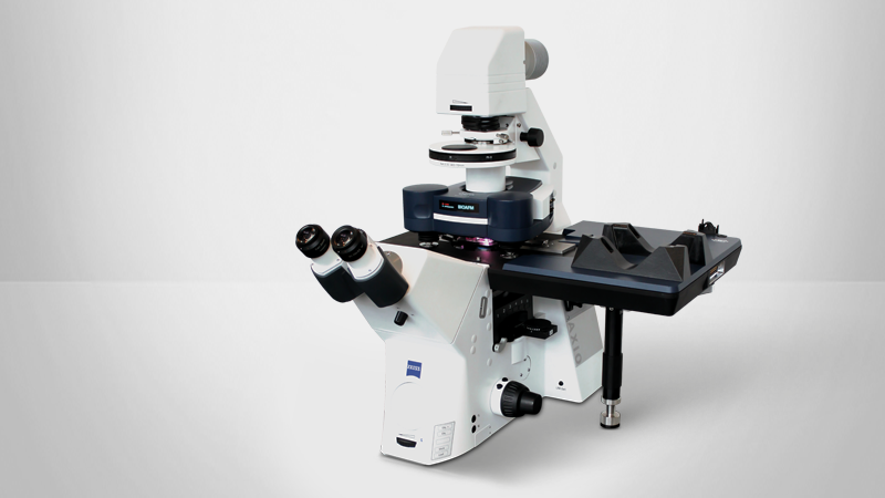
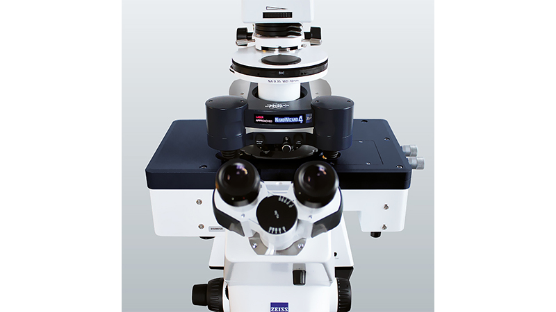
- Prof. Yakubov, from the School of Food Science and Nutrition, will discuss the BioFluidic Microscope (BFM), an integrated platform that combines confocal imaging with atomic force and nanofluidic functionality to explore biological and bioinspired systems in five dimensions.
Leading-Edge Research at the Intersection of Physics and Biology
Measurement of Plant Cells Turgor Pressure by AFM Indentation
Dr. Simoné Bovio, RDP laboratory and PLATIM-LyMIC imaging platform, Ecole Normale Supérieure, Lyon, France
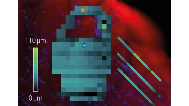
Plants cells can be imagined as balloons under pressure. Walled cells, as in plants, fungi or bacteria, can sustain a hydrostatic overpressure (turgor pressure) due to the osmotic pressure difference between the inside and outside of the cell. Turgor pressure (up to several MPa in plants) is the driving force of cell growth and participates in the overall mechanical stability of the plant. The interplay between turgor pressure and cell wall properties tailors cell growth (in rate and direction) and finally the overall morphogenesis (the generation of form) of a plant. Several approaches can be used to measure the turgor pressure (pressure chamber, psychrometry, pressure probe, etc.) that allow either an organ level or cell-level, but destructive, measurement. Furthermore, none of those techniques are adapted to small cells (below few tens of microns in radius). In this webinar, I will discuss the use of AFM indentation measurements to assess turgor pressure on a single cell level. I will present the different strategies developed in our lab and by other groups as well, highlighting their advantages and limitations. I will then show the results of an ongoing investigation on Physcomitrium patens phyllids.
Biofluidic Microscope: An Integrated Atomic and Fluidic Force – High-Resolution Confocal Microscopy Imaging Platform
Prof. Gleb E. Yakubov, School of Food Science and Nutrition, University of Leeds, United Kingdom
The BioFluidic Microscope (BFM) is a new, integrated imaging platform that combines ultra-fast confocal imaging with atomic force and nanofluidic functionality. The BFM enables the characterisation of localised biochemical and physiological processes across three dimensions of space, as well as time and force, thus offering a step-change capability for exploring biological and bioinspired systems in five dimensions: 3D space, time, and force (5D microscopy). The BFM unlocks new avenues for applications in soft matter, biomechanics, biomaterials, biofilm research, and the food and plant sciences. Its unique design allows it to accommodate a wide range of complex biological samples, enabling quantitative and predictive characterisation of single molecules, single cells, and tissues, as well as whole organisms or their larger fragments. The BFM’s applications span several domains, delivering benefits across a broad range of research areas, including biofilm dynamics, plant development, and cell physiology.
This talk will discuss the implementation of the BFM and illustrate its capabilities using two case studies: (a) nanomechanical mapping of A. thaliana roots and (b) interfacial interactions in plant-derived food materials. This will be followed by a discussion of approaches for the analysis of nanomechanical data in complex systems.
Featured Technologies:
Contact:
Find out more about the technology featured in this webinar or our other bioafm solutions: