Light-Sheet Imaging of Plants
Studying plants is immensely important to our understanding of biological mechanisms, facilitating crop improvement, and even ensuring global food security. Traditionally, plant samples were studied as thin dyed sections, but science is moving towards 3D in situ live imaging with technical advances in fluorescent tissue-specific reporter lines and genetically encoded biosensors. Imaging live plants in 3D comes with a set of challenges that require innovative solutions, and Bruker is uniquely qualified to address these challenges and experimental questions.
Challenges in Plant Imaging
One of the primary challenges of imaging plants is the inherent inhomogeneity of their tissues. Plant cells also have rigid cell walls and their chlorophyll absorbs light. Furthermore, plants exhibit natural tropisms with aerial parts oriented towards light and roots responding to gravity. These factors create the need for specialized sample setups that meet both physiological and physical conditions.
Plant Imaging Applications Include:
- Cytoskeletal biology and biophysics
- Cell cycle and death
- Phototropism and gravity
Using fluorescence microscopy for plant imaging is complicated by phototoxicity where lasers can affect plant health and function. Light-sheet fluorescence microscopy (LSFM) is an elegant solution to address this challenge of phototoxicity and is inherently less phototoxic than confocal imaging. LSFM systems also often offer the flexibility of vertical sample embedding and precise environmental control. Additionally, the thin light-sheet of SPIMs can penetrate through tissue and provide improved optical sectioning.
Live Imaging of Plants
Arabidopsis Root Growth
Transgenic Arabidopsis root expressing nuclear envelope marker imaged with the MuVi SPIM. The comparison of the videos shows the effect of gravity on root growth.
Courtesy of:
Shanjin Huang
Tsinghua University
Beijing, China
Arabidopsis Root
Transgenic Arabidopsis root expressing a membrane marker. Imaged with the InVi SPIM.
Courtesy of:
Alexis Maizel
COS, University of Heidelberg
Heidelberg, Germany

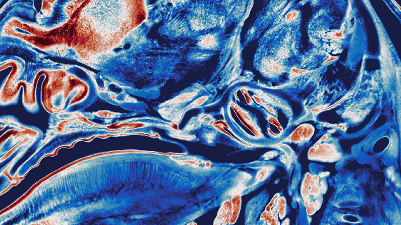
Microalgae
Autofluorescence in microalgae. Imaged on the InVi SPIM.