Application Note: Light-Sheet Imaging Reveals the Secrets of Neuroscience
Advanced Neuroscience Applications with Bruker’s Light-Sheet Microscopes
In this application note, readers can expect to learn about how light-sheet fluorescence microscopy (LSFM) overcomes traditional challenges in brain imaging by enabling high-resolution, low-phototoxicity visualization of complex neural structures across the brain, eye, spinal cord, and peripheral nervous system. Learn how the versatility of Bruker’s LSFM supports research in neurodevelopment, neurobiology, and disease modeling.
Contents include:
- LSFM addresses several brain imaging challenges
- Applications in neurodevelopment, neurobiology, and peripheral nervous system research
- Case studies using Bruker’s LSFM in mouse brain, zebrafish eye, and organoid models
- Comparison of Bruker LSFM systems tailored to specific neuroscience applications

The brain is highly complex and the most energy-demanding organ. It is composed of various cell types, including neurons that transmit information, immune cells that protect brain functions, astrocytes that regulate the chemical environment, and glia that provide structural and functional support. Additionally, non-cellular components, such as the cerebrospinal fluid (CSF) and (g)lymphatic system, are integral for maintaining brain health, homeostasis, and overall functionality. Due to its critical function, the brain, its health, and its development are major research areas.1 This application note discusses how light-sheet microscopy overcomes challenges in brain imaging to look at brain tissues and components, from the eye and organoids, to the spinal cord and peripheral nervous system.
Challenges in Brain Imaging
The brain is notoriously challenging to study. It is composed of countless small cells spread throughout a considerably large organ, often necessitating a trade-off between imaging the entire brain and achieving high-resolution images. For imaging in humans and other vertebrates, imaging at non-embryonic stages is challenging due to the skull encasing the brain with hard, opaque bone.
Advances in imaging correspond with changes in technology. In medicine, one of the gold standards for brain imaging in humans is functional magnetic resonance imaging (fMRI).2 The medical imaging landscape is rapidly changing because deep learning and artificial intelligence have a high impact on new developments for data acquisition and analysis.3 In contrast, for biomedical and preclinical models, changes in sample preparation techniques—such as brain expansion—often come into play. By physically expanding the brain tissue, the resolution issue can be overcome by increasing the sample rather than decreasing the resolution.
Another challenge in brain imaging is that in most organisms, the brain itself is not transparent but opaque, reducing light penetration and thus impacting imaging beyond the surface. To overcome this hurdle, tissue clearing can be used. Tissue clearing renders tissues transparent, enabling light penetration and imaging within the tissue. This allows the visualization of intricate neural networks and other brain structures.4

Neurobiology Versus Neurodevelopment
When it comes to brain research, two major areas of interest are brain development and neurobiology. Brain development studies the processes of brain formation and its maturation throughout life. It provides crucial insights into how abnormalities during development can lead to brain disorders.
Conversely, neurobiology is concerned with brain function, including memory, decision making, sensory perception, and more. This includes the understanding of neurotransmitters, neuroplasticity, and neurochemistry. Understanding neurobiology helps diagnose and treat various neurological conditions.

Light-Sheet Fluorescence Microscopy in Brain Imaging
Light-sheet fluorescence microscopy (LSFM) is a highly effective tool for brain imaging, offering practical advantages for neuroscientific research. Its notable features include penetration depth, reduced phototoxicity, and the ability to image fast processes.5 LSFM's versatility is evident in its applications for live imaging dynamic processes, such as calcium dynamics or long-term developmental changes in the brain. It is also ideally suited for imaging cleared and expanded tissues, offering cellular-level resolution of whole brains.6
Application Example—Hindbrain Morphogenesis
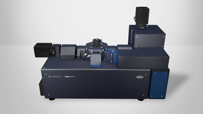
In one study, the role of Notch-3 in the neurogenic fate of hindbrain boundaries was described.7 The group used intricate time-lapse imaging and data analysis to show specific boundary cells during hindbrain morphogenesis depending on Notch-3 signaling. Bruker’s Multi-View Selective-Plane Illumination Microscope (MuVi SPIM) was employed for cell lineage analysis.
Imaging Beyond the Brain—The Eye
Eye research is essential to understand and prevent vision loss. Still, eye research is also often used as a proxy to understand the brain, as the eye is a peripheral part of the brain but with much greater accessibility. The eyes share numerous similarities with the brain regarding cell types, functionality, and cell-to-cell interactions. This structural and functional resemblance makes the eye an invaluable model for fundamental biological processes. Thus, eye research is not only critical in itself but also serves as a bridge to broader neurology.8
Application Example—Zebrafish Eye Laser Ablation
To understand macrophage response in the zebrafish eye, laser ablation can be used. Specifically, wounding of the eye is achieved using a laser, and a subsequent timelapse is acquired to understand macrophage recruitment and interaction with the wound. A transgenic line visualizing macrophages was used Tg(mpeg::gfp) with 488 nm excitation laser and collected with a 525/50 nm BP filter. Denoising and an unsharpen mask were applied to the maximum projection data. Each fish was a 150 µm volume acquired every 2 min.

Application Example—Subcellular Mouse Retina Dynamics
A group investigated mouse retinas in 3D using a MuVi SPIM to study subcellular Golgi apparatuses and tuft morphology.9


Imaging Beyond the Brain—The Spinal Cord
The spinal cord is central in transmitting signals between the brain and the rest of the body, influencing essential functions such as movement, sensation, and organ control. Advances in spinal cord research hold the promise of unlocking new treatments and therapies for spinal cord injuries, paralysis, and neurological disorders.10
Imaging the Peripheral Nervous System
By studying the peripheral nervous system (PNS), scientists gain unprecedented access to the intricate details of peripheral nerves. This not only aids in mapping the structural intricacies of nerve fibers but also allows for the visualization of dynamic processes crucial for understanding neural function. Biomedical imaging is an essential tool for elucidating the connectivity between the PNS and the brain, enabling the tracing of neural pathways and investigating how information is transmitted and processed. Moreover, studying the PNS is pivotal in diagnosing and monitoring neurological disorders that may manifest in both peripheral nerves and the brain.11 This integrated approach offers insights instrumental in advancing medical diagnostics, treatment strategies, and overall neurological research.

Cell Culture and Organoids
3D cell cultures are fragile systems that require optimized experimental protocols and low phototoxicity during imaging to preserve samples. Light-sheet imaging is a unique solution for these challenges by enabling researchers to study 3D cell culture systems, including time-lapse imaging.
Application Example—Astrocyte incorporation into neuronal organoids
A group studied how to establish human cell-based neural organoids and showed that the addition of astrocytes created a microphysiological system.12 Organoids were imaged with the MuVi SPIM microscope.

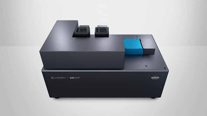
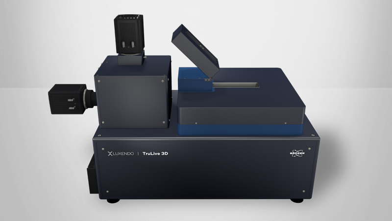
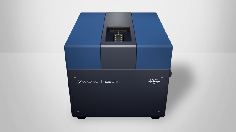
The Best Light-Sheet Microscopes for Different Neuroscience Applications
| Use Case | Scientific Requirements | Bruker |
| Large Cleared Sample |
| Large, cleared sample light-sheet fluorescence microscope |
| Fragile Samples |
| Dual-sided illumination light-sheet fluorescence microscope |
Live, Fixed, or Cleared Samples |
| Multiview imaging for live and cleared samples |
| Specialized High-Resolution Imaging |
| Inverted view light-sheet fluorescence microscope with advanced illumination |
Conclusion
The brain's role as the body's central control hub makes understanding its health paramount. Despite the challenges posed by its complexity, innovative techniques and approaches are continually advancing our understanding of brain structure and function, paving the way for breakthroughs in brain development and neurobiology research. This knowledge is crucial for improving the diagnosis and treatment of neurological disorders, ultimately enhancing the overall well-being of individuals.
Authors
- Dr Elisabeth Kugler, External Marcom Specialist, (E.kugler-extern@bruker.com).
References
- M. Bear, B. Connors, and M. A. Paradiso, Neuroscience: Exploring the Brain, Enhanced Edition: Exploring the Brain, Enhanced Edition. Jones & Bartlett Learning, 2020.
- R. A. Poldrack and M. J. Farah, “Progress and challenges in probing the human brain,” Nature, vol. 526, no. 7573, Art. no. 7573, Oct. 2015, doi: 10.1038/nature15692.
- M. Biswas et al., “State-of-the-art review on deep learning in medical imaging,” FBL, vol. 24, no. 3, Art. no. 3, Jan. 2019, doi: 10.2741/4725.
- T. C. Murakami et al., “A three-dimensional single-cell-resolution whole-brain atlas using CUBIC-X expansion microscopy and tissue clearing,” Nat Neurosci, vol. 21, no. 4, Art. no. 4, Apr. 2018, doi: 10.1038/s41593-018-0109-1.
- J. Huisken, J. Swoger, F. Del Bene, J. Wittbrodt, and E. H. K. Stelzer, “Optical sectioning deep inside live embryos by selective plane illumination microscopy,” Science, vol. 305, no. 5686, pp. 1007–1009, Aug. 2004, doi: 10.1126/science.1100035.
- M. B. Ahrens, M. B. Orger, D. N. Robson, J. M. Li, and P. J. Keller, “Whole-brain functional imaging at cellular resolution using light-sheet microscopy,” Nat Meth, vol. 10, no. 5, pp. 413–420, May 2013, doi: 10.1038/nmeth.2434.
- C. F. Hevia, C. Engel-Pizcueta, F. Udina, and C. Pujades, “The neurogenic fate of the hindbrain boundaries relies on Notch3-dependent asymmetric cell divisions,” Cell Reports, vol. 39, no. 10, Jun. 2022, doi: 10.1016/j.celrep.2022.110915.
- A. London, I. Benhar, and M. Schwartz, “The retina as a window to the brain—from eye research to CNS disorders,” Nat Rev Neurol, vol. 9, no. 1, Art. no. 1, Jan. 2013, doi: 10.1038/nrneurol.2012.227.
- C. Prahst et al., “Mouse retinal cell behaviour in space and time using light sheet fluorescence microscopy,” eLife, vol. 9, p. e49779, Feb. 2020, doi: 10.7554/eLife.49779.
- K. E. Lewis and J. S. Eisen, “From cells to circuits: development of the zebrafish spinal cord,” Progress in Neurobiology, vol. 69, no. 6, pp. 419–449, Apr. 2003, doi: 10.1016/S0301-0082(03)00052-2.
- J. Hubbard, The Peripheral Nervous System. Springer Science & Business Media, 2012.
- M. Brüll et al., “Incorporation of stem cell-derived astrocytes into neuronal organoids to allow neuro-glial interactions in toxicological studies,” ALTEX - Alternatives to animal experimentation, vol. 37, no. 3, Art. no. 3, Jul. 2020, doi: 10.14573/altex.1911111.
©2025 Bruker Corporation. InVi, TruLive3D, MuVi, and LCS are trademarks of Bruker. All other trademarks are the property of their respective companies. All rights reserved. AN2014, Rev. A0.