
Episode 2: Imaging Across Scales: From Subcellular Structures to Whole Organisms
Episode 2: Imaging Across Scales: From Subcellular Structures to Whole Organisms
In Episode 2 of The Light-Sheet Chronicles Podcast, host Dr. Elisabeth Kugler meets with special guests Dr. Urmas Roostalu (Gubra), Dr. Johanna Perens (Vibraint), and Bruker light-sheet microscopy expert Dr. Malte Wachsmuth to discuss applications from subcellular dynamics to whole-organ imaging, highlighting techniques like in vivo imaging, tissue clearing, and data analysis.
They address challenges such as data size, image tiling, and quality control. A standout moment features the "Doritos paper," showcasing how a food dye enables temporary tissue transparency in live animals.
Listen to this episode or scroll down to read more about the topics discussed.
Imaging Across Scales: From Subcellular Structures to Whole Organisms
Light-sheet fluorescence microscopy (LSFM) is more than a tool for generating stunning 3D images; it’s a way of uncovering biology across scales, from molecules and single cells to whole organs and organisms.
In the second episode of The Light-Sheet Chronicles, host Dr. Elisabeth Kugler is joined by three guests who bring different perspectives on light-sheet fluorescence microscopy:
- Dr. Urmas Roostalu: Senior Department Manager of 3D Imaging in biotech and drug discovery at Gubra, a company focused on preclinical drug development.
- Dr. Johanna Perens: Specialist in 3D imaging and image analysis at Vibraint.
- Dr. Malte Wachsmuth: Managing Director at Bruker Luxendo, the light-sheet division of Bruker.
Together, they explore how LSFM enables imaging across scales, how tissue clearing facilitates new insights, and how researchers are addressing the challenges of massive datasets.
Capturing In Vivo Dynamics
One of the main applications of LSFM is the study of development, or fast dynamic processes, such as understanding the morphogenesis of blood vessels in the trunk of zebrafish. What makes LSFM truly unique is its ability to capture fast biological processes in vivo with minimal phototoxicity.
As Malte Wachsmuth explains, this is crucial for studying subcellular dynamics, such as calcium signaling or analyzing blood flow: “Light-sheet microscopy lets us follow processes that happen on the millisecond scale, without destroying the sample in the process.”
Beyond rapid events, LSFM also supports long-term time-lapse imaging, for example, when studying morphogen gradients or organ development over hours and days. This is incredibly insightful, especially when combined with biomedical image analysis approaches, such as 4D cell tracking.
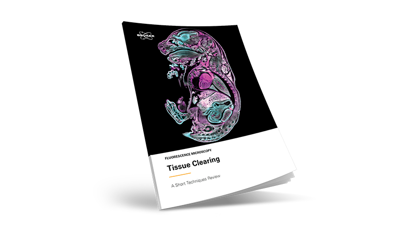
Tissue Clearing: From Preparation to Discovery
LSFM is also known to allow imaging across scales, from subcellular resolution to whole-organism datasets. This ability to image across scales is especially interesting when we want to understand, for example, the compound distribution of drugs at whole-organism levels. In many cases, imaging of large samples, such as whole organs or organisms, uses tissue fixation and clearing.1 Dr. Roostalu explains: “If we want to know how a compound distributes in an organ – say, whether it reaches fibrotic regions in the heart—we need whole-organ imaging. Clearing makes that possible.”

However, tissue clearing is not a one-size-fits-all technique. Different protocols balance speed, tissue preservation, and compatibility with labeling.
At Gubra, samples are often compared across treatment groups: “We can take several centimeters of tissue, image it at sub-micron resolution, and directly compare how drugs act in different organ regions.”
LEARN MORE ABOUT TISSUE CLEARING:
The Image Analysis Challenge
Of course, imaging across scales means generating large amounts of data. When acquiring full datasets of cleared organs, they often don’t fit within a single field of view (FOV) and thus require tiling and stitching, producing datasets that can be several terabytes in size.
Dr. Perens explains the computational side: “We rely on automation and standard atlas registration to make sense of these datasets. But at the end of the day, quality control and reproducibility still depend on careful human oversight.”
Here, collaboration across the community is essential, as biomedical image analysis workflows must evolve as a collaboration between biologists, microscopists, image analysts, and many others.2
The Doritos Paper: Transparency With a Twist
One of the most striking recent developments in the field is a paper that is often referred to as the “Doritos paper”, published in 2024.3 The study used tartrazine, a common food dye also found in Doritos, to temporarily make mouse abdomens transparent.
The science behind it comes from the Kramers–Kronig relation, where water-soluble dyes reduce refractive index (RI) mismatches between water and lipids.
The result: non-invasive, reversible transparency in living tissue.
From a photophysics perspective, tartrazine behaves like an electromagnetic oscillator: when excited near its resonance, only a small amount of energy is required to induce high-amplitude oscillations. Moving away from this resonance, however, higher energy input is needed, which has the side effect of increasing the refractive index in the corresponding wavelength range.
Tartrazine’s resonance occurs below approximately 450 nm, in the sub-blue and UV spectrum, and at sufficiently high concentrations, it increases the RI of aqueous solutions above this range. This is exactly what is needed to render live tissue temporarily transparent, as it raises the RI of cytoplasm and extracellular fluid closer to that of lipid- and protein-rich scattering structures, effectively reducing light scattering. This temporary effect enables imaging of live tissues previously only possible with fixed, cleared samples, opening the possibility of studying processes such as drug delivery to specific organs.
Malte Wachsmuth reflects on why this matters: “It sounds playful because of the Doritos connection, but it’s a serious breakthrough. If we can make tissues temporarily transparent without invasive clearing, it opens doors for live animal imaging that were previously closed.”
This discovery underscores how unexpected materials and cross-disciplinary ideas can reshape the field.
Imaging Across Scales in Practice
From subcellular calcium dynamics to fibrosis in the heart, LSFM shows its power in applications that demand imaging at multiple scales. Urmas Roostalu describes how a multiscale approach supports drug discovery by enabling the study of drug efficacy not only at the voxel level, but also across entire regions of an organ.
In the brain, for instance, this approach can reveal whether a compound engages circuits that regulate appetite, while simultaneously highlighting off-target effects, such as activation patterns that might trigger nausea, headaches, or migraine-like responses. These are insights that traditional, region-specific analyses simply cannot provide.4
Similarly, in kidney disease, particularly polycystic kidney disease, 3D imaging can capture the full landscape of cyst formation, growth, and distribution, showing precisely where a drug exerts an effect and where it does not.5
LEARN MORE ABOUT TISSUE CLEARING:
Wrapping Up
This episode of The Light-Sheet Chronicles highlights the breadth and depth of LSFM research, including:
- In vivo imaging captures fast dynamics and long timelapses with minimal damage.
- Tissue clearing enables organ-level studies of drug distribution and disease mechanisms.
- Image analysis remains a bottleneck, with terabyte-scale data pushing the limits of infrastructure.
- Innovative breakthroughs, like the “Doritos paper”, show that the field continues to surprise and inspire.
As light-sheet microscopy continues to evolve, it enables new science across scales.
References
[1] D. S. Richardson et al., “Tissue clearing,” Nat Rev Methods Primers, vol. 1, no. 1, p. 84, Dec. 2021, doi: 10.1038/s43586-021-00080-9.
[2] A. Schlaeppi et al., “Meeting in the Middle: Towards Successful Multidisciplinary Bioimage Analysis Collaboration,” Frontiers in Bioinformatics, vol. 2, 2022, Accessed: Apr. 19, 2022. [Online]. Available: https://www.frontiersin.org/article/10.3389/fbinf.2022.889755
[3] Z. Ou et al., “Achieving optical transparency in live animals with absorbing molecules,” Science, vol. 385, no. 6713, p. eadm6869, Sept. 2024, doi: 10.1126/science.adm6869.
[4] H. H. Hansen et al., “Whole-brain activation signatures of weight-lowering drugs,” Molecular Metabolism, vol. 47, p. 101171, May 2021, doi: 10.1016/j.molmet.2021.101171.
[5] U. Roostalu, H. H. Hansen, and J. Hecksher-Sørensen, “3D light-sheet fluorescence microscopy in preclinical and clinical drug discovery,” Drug Discov Today, vol. 29, no. 11, p. 104196, Nov. 2024, doi: 10.1016/j.drudis.2024.104196.


