Expanding your SEM Capabilities with Micro-XRF for SEM
Advancements in Microanalysis
An array of different analytical techniques exist to obtain geochemical information from small areas that are representative of single mineral phases. The most common analytical techniques are based on electron-beam (e-beam) sources, namely an electron probe microanalyzer (EPMA) or a SEM with an Energy Dispersive Spectrometer (SEM-EDS).
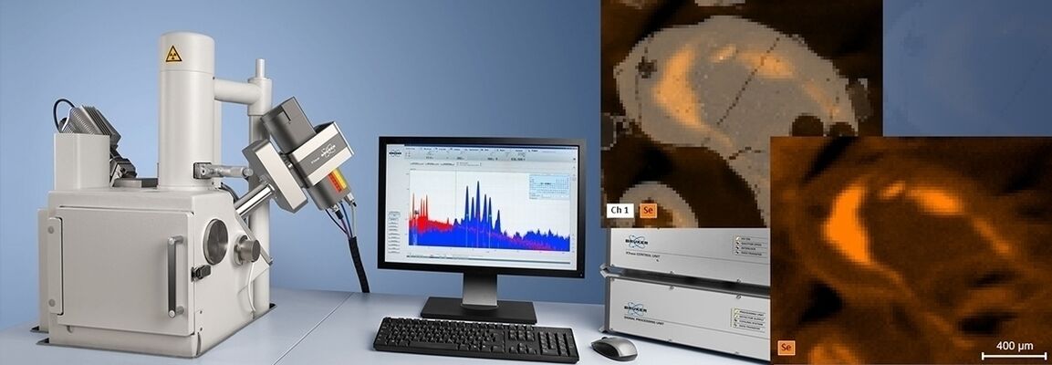
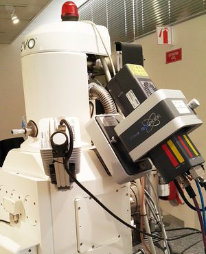
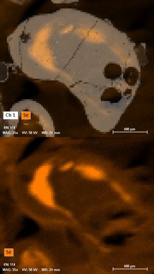
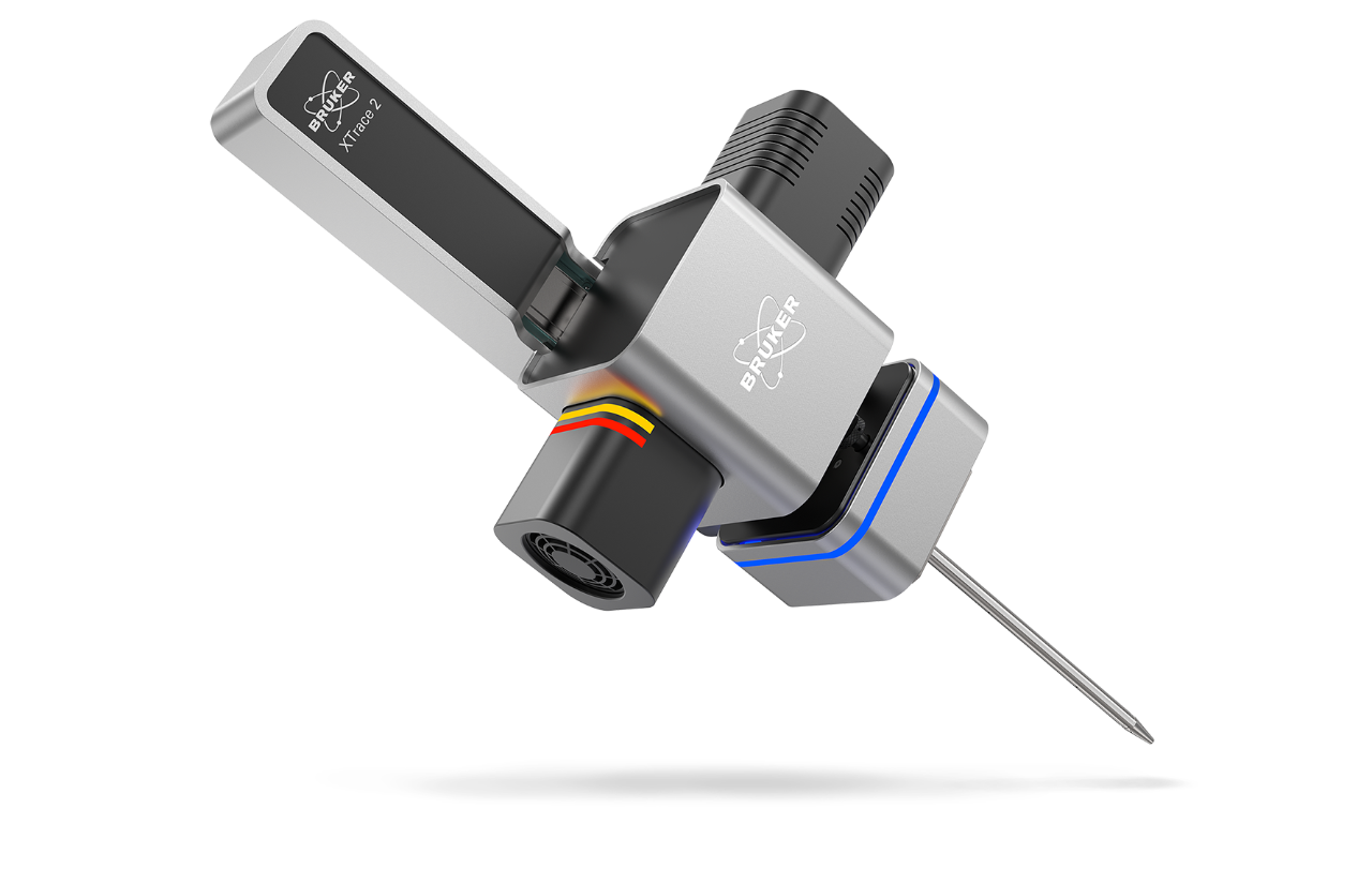
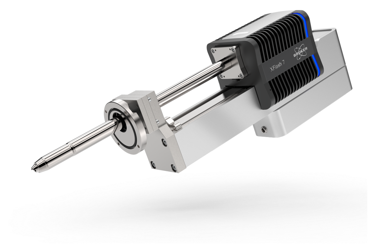
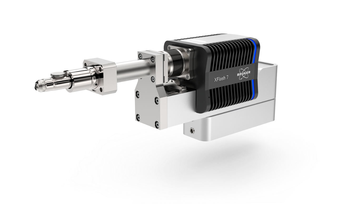
Similarly, Micro-XRF analysis can be considered as a complementary technique to e-beam excitation, which also delivers small area sample information. The Micro-XRF is based on photon excitation where samples will be directly excited with X-rays (X-Ray-beam), which has distinct differences compared to traditional e-beam based systems. The availability of modern X-ray polycapillary optics enables the capability to focus X-rays to a spot size of less than 30 µm, and all contained in the XTrace, a XRF source that can be mounted on an available SEM port (Figure 1). This results in a microscope system with dual beam potential i.e. both an e-beam and an X-ray beam. Accordingly, it is therefore possible to operate a Micro-XRF system using similar parameters as an e-beam system and thus yield results comparable with traditional point SEM-EDS analysis, whilst obtaining additional information from the X-Ray-beam sample interaction.
Each of the e-beam and X-Ray-beam techniques have implicit advantages and limitations, especially with respect to analytical volume and detection limits, as well as sample interaction. This webinar compares these different techniques with regard to spatial resolution and sensitivity for point analysis, line scans and area maps using a variety of geological examples (e.g. volcanic specimens (Figure 2), mantle xenoliths and xenocrysts, meteorites, Exotic-Cu deposits, amongst others to demonstrate how Micro-XRF combined with a SEM-EDS extends the analytical capabilities of the system.
This webinar will conclude with a 15-minute Q&A session where our experts will answer your questions.
Who Should Attend?
- Researchers in Geological Sciences and Mining
- Scientist from analytic fields interested in Micro-XRF technology for SEM´s
Speakers

Andrew Menzies, Ph.D.
Universidad Católica del Norte (Chile) Department of Geological Sciences

Stephan Boehm
Product Manager - micro-XRF on SEM and WDS, Bruker Electron Microscope Analyzers
Watch this Webinar On-Demand
Please enter your details below to gain on-demand access to this webinar.