Secondary Structure of Proteins and Peptides
Secondary Structure of Proteins and Peptides
Circular dichroism is known for the ultraviolet (UV) spectral region. Applications are limited in most cases because of characteristic absorption bands are typically broad and superimposed by unspecific bands (e.g. aromatics side chains). Especially for biomolecules large absorption and dispersion coefficients limit the applications.
In the infrared (IR) molecular vibrations are excited. The according absorptions bands are narrow and specific in general. A masking of the information unlike in electronic CD is not observed in the IR.
The following figure shows VCD (upper) and absorption (lower) spectra of hemoglobin (left), lysozyme (mid) and concanavalin A (right) each solved in D2O solution (50 mg/ml) in a 25 µm CaF2 cell.
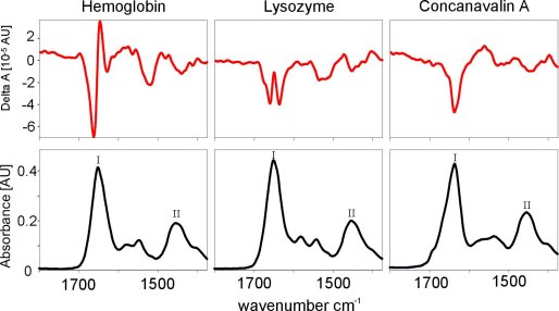
The absorption spectra for the three proteins are very similar. All show a comparatively broad unstructured amid I absorption band (mainly C=O stretching mode), indicated by "I", and amid II absorption band (mainly C-N stretching and N-H deformation mode), indicated by "II".
First at a closer examination it is apparent that the amid I band of the hemoglobin behaves asymmetric around 1650 cm-1 while concanavalin A exhibits its amid I absorption maximum at 1630 cm-1 and a small shoulder at 1690 cm-1.
Despite of similar absorption spectra these proteins show significant differences in their secondary structure: hemoglobin is determined by an α-helix structure mainly while concanavalin A exists in a β-sheet predominantly and lysozyme shares both structure elements in comparable amounts.
Much more apparent are the differences for the secondary structures in VCD spectra. The derivate-like band of haemoglobin is for a high α-helix contribution typical whereas the dip at 1630 cm-1 in the VCD spectrum of concanavalin A is very specific for β-sheet structures. So it is no surprise that the VCD spectrum of lysozyme consists of both structure elements.
VCD spectroscopy therefore provides a unique possibility to determine the secondary structure of proteins but allows for the observation of conformational changes also. Experiments on other proteins show that signals in VCD depend from secondary structures unambiguously.


