RAMANtouch
Use the Line.
And Stop Wasting Time.
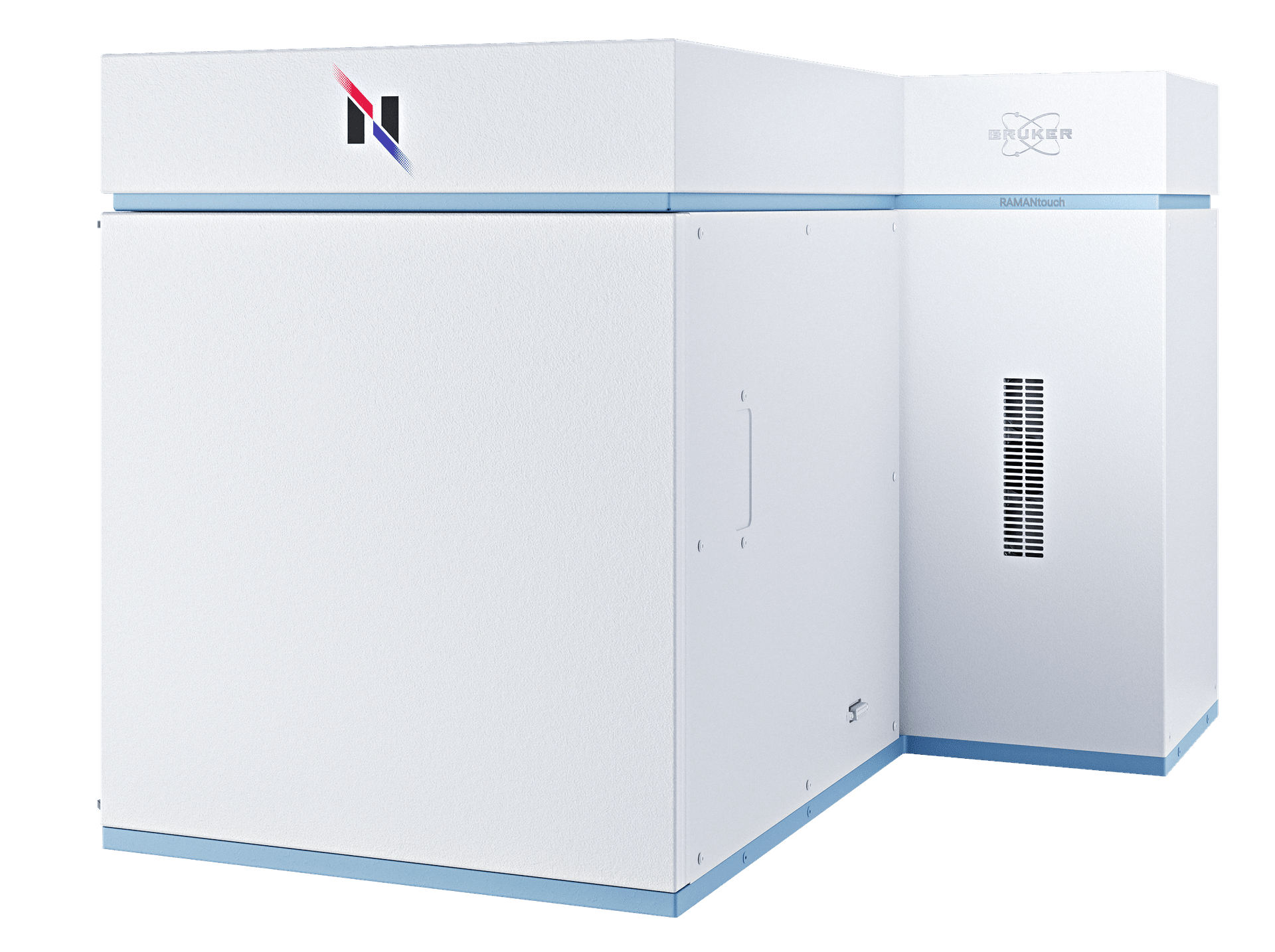
A confocal Raman imaging microscope with unsurpassed speed and accuracy.
Nanophoton's tech uses line-shaped laser illumination and a two-dimensional CCD to capture 400 spectra in one exposure. With laser scanning via galvanometer scanners, RAMANtouch achieves rapid and accurate Raman imaging of hundreds of thousands of pixels within minutes - no EMCCD required.
True Raman imaging is finally here.
The RAMANtouch is a completely new approach compared to traditional Raman microscopes - and that's why it achieves the impossible. Maximum imaging speed without any loss of spectral quality or spatial resolution.
The RAMANtouch offers:
- Ultra high-speed Raman imaging
- High spectral and spatial resolution
- Completely automated hardware
- Powerful analysis for 2D/3D Raman images
- Auto-calibration and auto-alignment
The Best RAMANtouch Features and How They Help You.
Discover the Nanophoton Technology
Discover how the Nanophoton RAMANtouch transforms Raman microscopy with innovative galvano-scanning technology. From precise point measurements to high-speed line scans and rapid large-area imaging, RAMANtouch offers the flexibility to reveal chemical information faster and more efficiently than conventional approaches.
Its unique measurement modes are designed to help researchers capture more data in less time while maintaining exceptional spatial resolution and image quality. Whether you're exploring advanced materials, batteries, semiconductors, or pharmaceuticals, RAMANtouch opens new possibilities for Raman imaging and analysis.

Exceptional 3D Raman imaging capabilities.
Confocal optics allow non-destructive Raman analysis inside a sample. This way, 3D Raman images of transparent samples can be created. By using line illumination and the high-precision stage of the RAMANtouch, 3D Raman images are created in unmatched speed and quality to provide insight into a sample's internal structure and component distribution.
Video example:
- Raman detects inside the transparent fiber
- Sample: Bi-component PE (cladding) and PET (core) fiber
- Acquisition time: 20~30 min
- Yellow dots: TiO2
Peak Raman imaging resolution.
Depth profiling Raman imaging in the XZ plane enables non-destructive analysis, such as examining multilayer films. Utilizing an oil-immersion objective lens significantly enhances spatial resolution, allowing for the detection of ultra-thin layers as fine as 250 nm.

Access the nanometer domain.
Raman microscopy offers essential insights into the identification of both organic and inorganic compounds, as well as crystal polymorphism. Unlike IR microscopy, Raman boasts advantages such as sub-micrometer spatial resolution and the capability to analyze samples in aqueous environments.


RAMANtouch specifications.
| Spatial Resolution | 350 nm in X, 500 nm in Y; 1 µm in Z | |
| Objective Lenses | 5x, 10x, 20x, 50x, 100x | |
| Spectral Resolution | <0.9 cm-1 (depends on grating, up to 3 gratings available) | |
| Stage Details | 30 * 30 * 35 mm XYZ-motorized stage | |
| Calibration | Auto-calibration based on standard lamp and sample | |
| Alignment | Auto-alignment of optical path | |
| Laser Safety | Laser safety class I door with interlock |

When I first encountered a Bruker’s Nanophoton Raman microscope, which was installed for the first time at Analytical center in Chungbuk National University, I was very surprised by the various performances that are different from the previous Raman microscopy. In particular, I was very impressed the function of large-area mapping, which is a disadvantage of most of current Raman microscopy, was solved with the idea of line-illumination.
In our laboratory, Raman spectroscopy was utilized to quantify amino acids in renal cell cultures from an animal model of induced renal dysfunction and to study the distribution of drugs on the cell membrane surface. In addition, the content of illicitly distributed APIs-like compounds was determined by Raman imaging and a semi-quantification method was established.
In addition, the bio-samples that we are mainly researching in our laboratory are very difficult to find the optimum experimental conditions due to the control of the laser power, but the RAMANtouch model can not only adjust a very small power level, but also use the preview function to find the optimal conditions faster than any other Raman product. By using such a powerful Raman microscopy in our laboratory, we have published several number of related papers.
Professor Dr. Yong-Moon Lee, School of Pharmacy, Chungbuk National University, Korea.

The RAMANtouch microscope has made us rethink the limits of application of Raman imaging. The multiplexed acquisition approach brings a significant increase in speed, making it feasible to collect large Raman images instead of few measurement points. The system automation enables our users to utilize measurement time efficiently in overnight runs. This capability adds analytical depth to Raman characterization and facilitates a better understanding of complex research questions and enables big data exploration with machine learning tools.
At Synchrotron Soleil, we facilitate scientific progress by providing access to a high level research infrastructure. Beyond the beamline instrumentation, we also provide additional characterization tools such as offline Infrared or Raman microscopy.
Ferenc Borondis, Beamline Manager & Principal Beamline Scientist at Synchrotron SOLEI, Saint-Aubin, France.

Now, with the pioneering application of the modifed RAMANtouch system to the world’s first 8.5th generation display mass production line, we can achieve groundbreaking real-time defect analysis directly within the production process. By enabling component-level analysis of random defects—responsible for over 90% of yield-loss issues—this technology allows for precise defect identification, helping manufacturers not only trace defect origins and specify affected processes but also prevent these issues in real time.
In the high-stakes realm of display product mass production, advanced inspection technology has become essential for optimizing yield and ensuring top-tier quality control. As these technologies evolve, the need for faster, more precise process feedback has driven demand for real-time monitoring that goes beyond traditional inspection to include advanced measurement and in-depth analysis. Furthermore, In-line customized RAMANtouch’s proactive feedback capabilities offer unprecedented opportunities for continuous process improvement and accident prevention, marking a transformative step forward in display production technology.
Dr. Yong-Woon Lim, Metrology & Inspection Team, Samsung Display, Korea.
Frequently Asked Questions
- Why does RAMANtouch achieve high spatial resolution?
RAMANtouch is designed strictly according to optical principles and assembled by specialists in optical design, which ensures that every unit achieves stable and reproducible performance. Its compact mechanical structure also minimizes the likelihood of misalignment caused by environmental changes. - Do I need to adjust the optics when switching lasers?
No manual adjustments are necessary. When a laser is switched, the required optical adjustments are performed automatically, and the change can be carried out with a single click. - Do I need to adjust the optics when switching diffraction gratings?
No. Just like laser switching, the instrument automatically adjusts the optics after the grating is changed, and the user does not need to perform any manual alignment. - Why are line illumination and laser beam scanning used?
These methods reflect Nanophoton’s origins in laser microscopy. Laser beam scanning eliminates the need to adjust measurement positions by moving a motorized stage and avoids issues such as vibration or sample displacement introduced by stage movement. The proprietary line illumination method enables the acquisition of 400 Raman spectra in the X-direction with a single irradiation, allowing high-speed Raman imaging. - How is illumination unevenness in line illumination addressed?
Although uneven intensity between the center and edges of the line is a common concern in line-illumination systems, RAMANtouch significantly reduces this issue using a proprietary technology covered by U.S. Patent US7561265. - Can high-power laser irradiation damage samples?
RAMANtouch includes an ND filter that provides nearly stepless control of laser power across 256 levels. This allows high power to be used efficiently for samples that tolerate it, while also enabling safe measurement of heat-sensitive samples by reducing the power appropriately. - How large is the field of view?
The field of view depends on the objective lens. With a 20× objective, the observation area is about 400 µm × 600 µm and the maximum imaging range is approximately 400 µm square. With a 100× objective, the observation area is roughly 80 µm × 100 µm and the maximum imaging range is about 80 µm square.