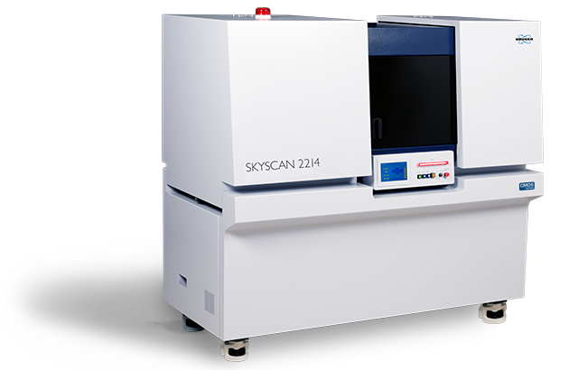
SKYSCAN 2214 CMOS EDITION
Ultimate Performance at your Command
Highlights
SKYSCAN 2214 CMOS EDITION– Nanoscale X-ray Microscopy
The new MULTISCALE X-ray nanotomograph SKYSCAN 2214 CMOS EDITION covers the widest range of object sizes and spatial resolutions in a single instrument. It opens unique possibilities for 3D imaging and exact modeling of all types of biological samples, including mineralized tissues, soft tissues, plants, insects, paleontological samples, etc.
The instrument allows scanning and 3D non-destructive reconstruction of the internal microstructure of objects as large as 300mm in diameter as well as submicron resolution for small samples.
The system contains an "open type" transmission X-ray source with <0.5 microns spot size and a diamond window. It can be equipped with up to four different X-ray detectors for maximum flexibility: flat-panel for large objects, 16Mp CMOS with large field of view, 16Mp cooled CMOS with standard field of view, and 15Mp cooled CMOS for highest spatial resolution. Automatic variable acquisition geometry and phase-contrast enhancement allows best possible quality in relatively short scanning time.
The SKYSCAN 2214 CMOS EDITION is complemented by 3D.SUITE. This comprehensive software suite covers GPU-accelerated reconstruction, 2D/ 3D morphological analysis, as well as surface and volume rendering visualization.
What is XRM? video
Key Features
Source
The SKYSCAN 2214 CMOS EDITION uses a latest generation open-type X-ray source. The source offers true spatial resolution below 500 nm, an X-ray energy up to 160 keV and source power up to 16 W. The source is practically maintenance-free with an extremely easy pre-aligned filament replacement procedure.
The SKYSCAN 2214 CMOS EDITION has an open-type (pumped) nanofocus X-ray source with diamond window. It produces an X-ray beam with peak energy from 20 kV to 160 keV and is supplied with two types of cathodes. The tungsten (W) cathodes operate in the full range of accelerating voltages up to 160 kV and provide a spot size down to 800 nm. The lanthanum hexaboride (LaB6) cathodes can be used for accelerating voltages from 20 kV to 100 kV and provide a spot size of the X-ray beam smaller than 500 nm to achieve the highest resolution in imaging and 3D reconstruction. The JIMA resolution pattern indicates that 500 nm structures can be easily resolved.
For long-term stability of the focal spot size and position of the emission point, the X-ray source is equipped with a liquid cooling system which contains a re-circulator providing precise temperature stability of the cooling fluid.
Detectors
The SKYSCAN 2214 CMOS EDITION can be equipped with up to four X-ray cameras for ultimate flexibility: three CMOS cameras with different resolution and field of view and one large-area flat panel detector. All cameras can be selected with a single mouse click. The different CMOS cameras can be retro-fitted at any point of time during the system’s lifetime.
All three CMOS can take images in the central beam position and in two offset positions to double the field of view. The images in the two offset positions are automatically stitched together with compensation of the shifts and possible intensity differences.
Using CMOS detectors with small pixel size allows extension of high-resolution imaging and 3D reconstruction to large objects. The built-in detector flexibility enables adjusting the field of view and spatial resolution according to the object size and density. An advanced reconstruction from a volume of interest provides scanning of a selected part of a large object with high resolution without compromising image quality.
Additionally, the field of view can be increased horizontally and vertically by using offset camera positions and vertical object movement.
In-situ stages
The high-precision object stage of the SKYSCAN 2214 CMOS EDITION supports objects up to 300 mm diameter and 20 kg in weight. The air-bearing rotation motor allows precise rotation of objects at very high accuracy, and the integrated micro-positioning stage guarantees a perfect sample alignment.
The SKYSCAN 2214 CMOS EDITION has a large and easily accessible sample chamber to allow scanning of big objects as well as mounting of optional stages. On top plenty of space is available for peripheral equipment.
The Bruker material testing stages are designed to perform compression experiments up to 4400 N and tensile experiments up to 440 N. All stages automatically communicate through the system’s rotation stage, without the need of any cable connections. Using the supplied software, scheduled scanning experiments can be set up.
Bruker's heating & cooling stages can reach temperatures of up to +80 °C or 30 °C below ambient temperature. Just like the other stages, no extra connections are needed, and there is an automatic recognition of the stage. Using the heating & cooling stages, samples can be examined under non-ambient conditions, to evaluate the effect of temperature on the sample’s microstructure.
The SKYSCAN 2214 CMOS EDITION is fully compatible with stages from DEBEN. With the included adapter, the DEBEN stage can be simply placed onto the rotation stage of the SKYSCAN 2214.


Highlighted Applications
Mineralized Tissues

The SKYSCAN 2214 CMOS EDITION provides ultimate nanoCT imaging performance without compromise. Four cameras are fitted – one large format 6 megapixel cMOS flat panel for imaging up to high voltages and large skeletal or dental samples, and a range of three small format detectors for the optimal choice of resolution and x-ray energy window for every conceivable bone sample scan.
Submicron voxel scans provide crystal clear resolution of bone osteocyte lacunae and micro-scale mineralization structure, as well as architecture of biomaterial scaffolds at nano-scale resolution.
Phase retrieval (Paganin) provides a new dimension of analysis of hitherto little studied bone micro-mineralization patterns.
Orthopedic research with sheep, primate or similar models is handled with the 14cm scan field of view and 160kV x-ray source. Realize your orthopedic and biomechanical research goals with mechanical testing and temperature control stages.
Morphometry with comprehensive 3D and 2D parameters, with densitometry including BMD calibration references over the preclinical size range. Advanced 3D image analysis functions include 3D registration, adaptive thresholding, Euler connectivity, fractal, anisotropy and stereology, filtering, Boolean logical operators and much more.
Soft Tissues

Ex vivo nano-CT scanning of biological tissues, with sub-micron pixel size, is like histology or electron microscopy but in true, deep 3D - a fantastic method to show internal structures non-destructively. Contrast agents or chemical drying can improve the image quality by further enhancing or differentiating tissue densities. The SKYSCAN 2214 CMOS EDITION is playing a leading role in the emergence of a new imaging discipline - microCT histology and histomorphometry.
The unmatched versatility of this system ensures that each sample will be scanned with optimized parameters and the best resolution and contrast.
Four cameras are fitted - a large format 6 megapixel flat panel for imaging up to high voltages and large tissue samples, and a range of three small format cameras for the optimal choice of resolution and x-ray energy window for every conceivable sample scan.
Comprehensive 3D image analysis capability including morphometry and densitometry, 3D registration, segmentation and advanced image processing methods.
Plant and Animal Biology

MicroCT is exceptionally good for the visualization of the finest details of internal biological structures. This imaging method creates a nano-scale 3D x-ray attenuation map without harming or destroying the scanned object. Practically all biological tissues can be visualized and analyzed with little or no need of special sample treatment. Submicron voxel resolution allows imaging of small insect and plant or seed structures with powerful magnification and rich detail.
The space in the scanning chamber and the precision of the rotation stage make sure that all types of samples can be scanned, from the small zebrafish and preserved zoological and botanic samples to potted plants and rock-embedded fossils.
Four cameras are fitted – a large format 6 megapixel flat panel for imaging up to high voltages and large samples, and a range of three small format cameras for the optimal choice of resolution and x-ray energy window for every conceivable biological sample scan.
The software suite enables morphometry with comprehensive 3D and 2D parameters, and advanced 3D image analysis functions include 3D registration, adaptive thresholding, Euler connectivity, fractal, anisotropy and stereology, filtering, Boolean logical operators and much more.
Application Gallery
3D model of an entire bee head (left), and virtually cut to expose internal structures (right), scanned at 1.4 µm voxel size.

3D model of a toothpick, scanned at nanoscale resolution.

Volume rendered image of a subvolume of dentin from a human molar, scanned at 350 nm voxel size. The model is virtually cut to show the dentine tubules running through the dentin in blue.

Orthogonal slices though a mouse distal tibia showing the osteocyte lacunae, scanned at 1 µm voxel size.

Volume render of a piece of wood, scanned at 200 nm voxel size (left) with the vascular network color-coded for local thickness (right).

3D model of a sheep bone with 2 large titanium implants.

Mouse lungs scanned at 3 µm voxel size after chemical drying.

3D Volume rendered image of a medieval human skull from Thuringia, scanned at 50 μm voxel size (Sample from the Friedrich-Schiller University Jena, Germany).

Cross-section through a Brassica seed, scanned at nanoscale resolution.

SKYSCAN 2214 CMOS EDITION Specifications
| Feature | Specification | Benefit |
| X-ray source | 20-160 kV 16 W max. | User exchangeable filament Optimize for max power (W) or max resolution (LaB6) Rotatable diamond window for maximum lifetime |
| X-ray detector | 6 Mp active pixel flat-panel 16 Mp large field of view CMOS 16 Mp standard field of view CMOS 15 Mp high resolution CMOS | Variety of pixel and detector sizes allow balance between detector resolution, coverage and counting statistics Available with 1, 2, 3 or 4 detectors Field upgradable to add detectors |
| Image Formats | Up to 8000 x 8000 x 2300 pixels after a single scan | User selectable image size allows balance of dataset size and needed resolution Software allows downsizing after data collection |
| Resolution | 60 nm smallest pixel size <500 nm low-contrast resolution (10% MTF) | Simple graphics control for optimizing experiment resolution based on selected detector, sample and detector distance Adjustable source focus size to balance maximum power and resolution |
| Positioning Accuracy | <50 nm for rotation Anti-vibration granite platform with pneumatic leveling | Air bearing sample stage provides smooth rotation Simple chuck style mounting of sample posts Mechanical and electrical interface for advanced materials research stages |
| Maximum Object Size | 300 mm in diameter (140 mm scanning size) | Both the power and room to scan large samples |
| 400 mm in length | Precise positioning of small samples near the source to maximize magnification | |
| Maximum object weight 20 kg | ||
| Dimensions | W 1800 mm x D 950 mm x H 1680 mm Weight: 1500 kg | Efficiently designed to optimize use of lab space Source maintenance access via interlocked large sliding door |
POSITION, SCAN, RECONSTRUCT and ANALYZE

Bruker XRM solutions include all software needed to collect and analyze data. An intuitive graphical user interface with user guided parameter optimization support both expert and novice users. By using the latest GPU powered algorithms, reconstruction time is substantially reduced. CTVOX, CTAN and CTVOL combine to form a powerful suite of software for both qualitative and quantitative analysis of models.
3D.Suite Software:
Bruker microCT solutions include our comprehensive, in-house developed
3D.SUITE software for reconstruction, inspection, visualization, and
analysis of the internal object structure
Measurement Software:
SKYSCAN 2214 – Instrument control, measurement planning and collection
Reconstruction Software:
NRECON – Transforms the 2D projection images into 3D volumes
Analysis Software:
DATAVIEWER – Slice-by-slice inspection of 3D volumes and 2D/3D image registration
CTVOX – Realistic visualization by volume rendering
CTAN – 2D/3D image analysis & processing
CTVOL – Visualization of surface models to export for CAD or 3D printing
SKYSCAN 2214 CMOS EDITION - Service & Support
Available services include
- Help desk support from highly-skilled troubleshooting professionals, to isolate and resolve hard- and software problems
- Web-based remote instrument service for service diagnosis and applications support
- Merged reality support with Help Lightning – a virtual engineer at your side (video)
- Planned maintenance, according to your requirement
- Customer on-site repair and maintenance service
- Spare parts availability typically over night or within a few working days worldwide
- Compliance services for installation qualification, operational qualification / performance verification
- Site planning and relocation
Bruker Support
On our support website you will find:
Software Updates
- System software updates are available online to registered users.
Documentation
- Product manuals and installation guides are available online to registered users.
LabScape
Service & Life Cycle Support for Magnetic Resonance and Preclinical Imaging
Bruker’s commitment to provide customers with unparalleled help throughout the buying cycle, from initial inquiry to evaluation, installation, and the lifetime of the instrument is now characterized by the LabScape service concept.
LabScape Maintenance Agreements, On-Site On-Demand and Enhance Your Lab are designed to offer a new approach to maintenance and service for the modern laboratory