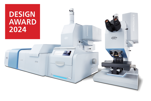
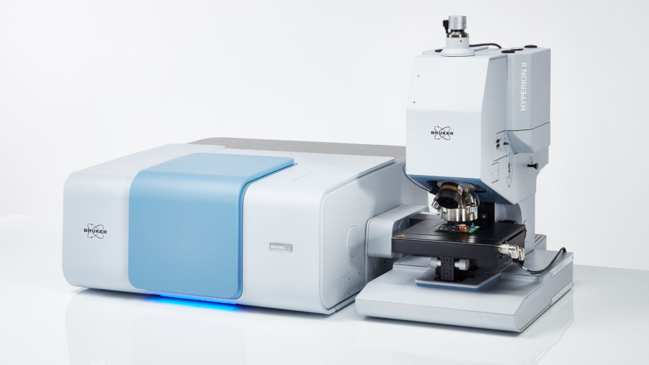
HYPERION II
FT-IR meets QCL
Analytical Power and Innovation
The IR Microscopy and Imaging Research Platform
What's new with HYPERION II:
How we integrated infrared laser imaging:
Our patented spatial coherence reduction:
FT-IR Microscopy Augmented by QCL | Infrared Laser Imaging
The HYPERION II is an innovation force in infrared microscopy. It provides IR imaging down to the diffraction limit and sets the benchmark in ATR microscopy. It combines FT-IR and Infrared Laser Imaging (ILIM) microscopy for the first time ever in a single device, offering all three measurement modes: transmission, reflection, and ATR.
HYPERION II features:
- Selection of detectors for µ-FT-IR:
Broad-, mid, narrow-band LN2-MCTs,
thermoelectrically cooled (TE) MCT. - Focal-plane array detector for infrared imaging (64 x 64 or 128 x 128 pixel).
- Optional QCL implementation by Laser Infrared Imaging Module (ILIM, laser class 1)
- Objective lens selection: 3.5x/15x/36x IR,
20x ATR, 15x GIR, 4x/40x VIS. - Spectral range extension – from Near-Infrared (NIR) to Far-Infrared (FIR)
- Selection of apertures: manual knife-edge, automated knife-edge aperture wheel. Metal apertures for NIR
- Selection of accessories and sample stages: macro IR imaging accessory, cooling/heating stage, sample compartment, etc.
- Selection of visual/optical tools: Darkfield illumination, Fluorescence illumination, VIS polarizers, IR polarizers, etc.
HYPERION II provides:
- Perfect match of spectral and visual images. Applies to any measurement mode (including ATR imaging).
- Diffraction limited high sensitivity FT-IR microscopy and imaging by using focal plane array (FPA) detector.
- First ever combination of FT-IR and QCL technology by (optional) Infrared Laser Imaging Module (ILIM, laser class 1).
- Infrared laser imaging in all measurement modes (ATR, Transmission, Reflectance).
- Patented coherence reduction for artifact free Laser Imaging measurements without sensitivity or speed loss.
- High imaging speeds:
0.1 mm2per second (FPA, full spectrum)
6.4 mm2 per second (ILIM, single wavenumber) - Optional TE-MCT detector to perform IR microscopy with high spatial resolution and sensitivity without liquid Nitrogen.
- Emission spectroscopy capability and optional spectral range extensions.
HYPERION II applications:
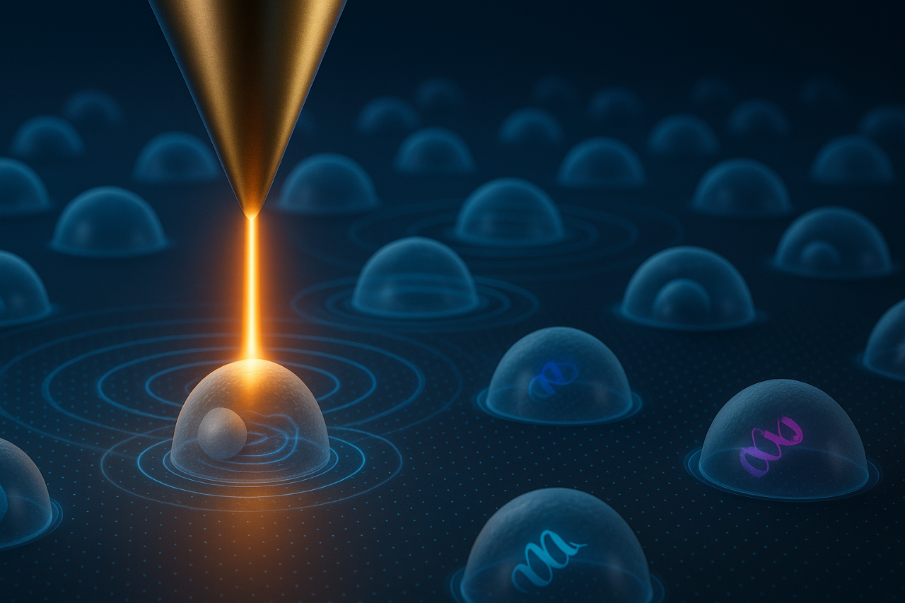
- Life science | cell imaging
- Pharmaceuticals
- Emissivity studies (e.g. LEDs)
- Failure and Root Cause Analysis
- Forensics
- Microplastics
- Industrial R&D
- Polymers & Plastics
- Surface characterization
- Semiconductor
The FT-IR Research Microscope for Pioneers and Innovators
Hardly any of our IR microscopes embody our users like the HYPERION II:
Flexible, precise, configurable, adaptable, and always at the limit of what is possible.
Take Full Control
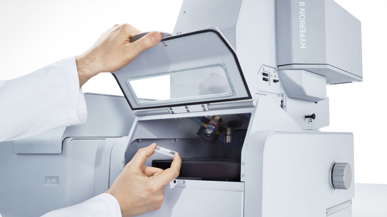
Above all, it is about having complete access to an instrument. Access to the experiment, the samples, and the parameters. This is the foundation of the HYPERION II and its most valuable asset: providing full control.
Whether FT-IR measurements in single point mode, mapping or imaging with different detectors or objectives, special sample stages or with ATR or Grazing Angle objective. At any point you can influence the outcome of your results - and make them better.
This is the clear difference to our LUMOS II IR microscope. Where the LUMOS II relieves the user of tedious experimental details and automates the measurement process, the HYPERION II remains a precise tool that only does what the user demands.
A Monument to its Past
Many users know the HYPERION II and its strengths through its predecessor. For almost 20 years, it has been an innovation force in IR microscopy and imaging. The things that made HYPERION an outstanding FT-IR microscope are still there - only better, faster and improved.
The HYPERION II continues to have all the features you require in your daily research routine: liquid nitrogen and thermoelectrically cooled MCTs, focal plane array imaging detectors, visual and infrared enhancement tools, and of course an abundance of dedicated accessories.
In the end, we wanted to set the benchmark in FT-IR microscopy and imaging once again and live up to our name as an innovation leader by introducing new and exciting technology while keeping established and valued methods.

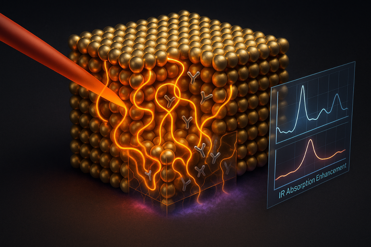
Augmenting FT-IR by Infrared Laser Imaging (QCL)
QCL and FT-IR in a single instrument
For the first time, users can access an IR microscope that combines FT-IR and QCL technology in one instrument. With this, we are opening a completely new door to life science and material research.
Collect an FT-IR spectrum, select the wavelengths you want to investigate using QCL and create stunning chemical images in a matter of seconds.
With this completely new approach of FT-IR and infrared laser imaging, we finally give users, researchers, and scientists a tool to develop new applications, but also to improve established and proven approaches.
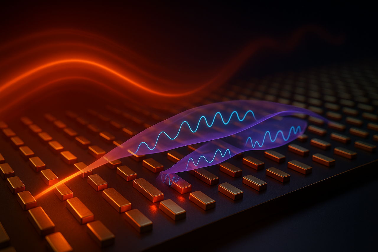
A true QCL microscope with exceptional performance
The HYPERION II offers uncompromised QCL microscopy in a state-of-the-art FT-IR microscope. In fact, we have specifically developed and patented a novel coherence reduction technology to enable unparalleled IR laser imaging performance - without digital post-processing.
To illustrate: In classical FT-IR, spatial coherence does not play a role. In IR microscopic measurements with a QCL, however, spatial coherence phenomena inevitably occur. These fringes and speckles in IR images and spectra are generally considered to be harmful for chemical imaging (see adjacent; DOI: 10.1002/jbio.201800015).
Indeed, it is not trivial to separate the sample's chemical information from the physical information describing the phase relationship of the scattered photons. The HYPERION II addresses this problem pragmatically and solves it by smart hardware design and lets you acquire artifact-free chemical imaging data.
Comparing FT-IR and QCL spectroscopy
Comparing the techniques would imply that both can perform the same task equally well - a popular misconception. FT-IR and infrared laser imaging have distinct advantages and only a practical combination of both can achieve the best results.
We know that most scientists and researcher do not want to miss the universality of FT-IR. They don’t like being restricted to a single, cutting-edge technique without point of reference. Fortunately, the HYPERION II can be considered both: an exceptional FT-IR imaging microscope and an ambitious QCL microscope.
We have addressed this duality and where QCL technology records data significantly faster at the same signal to noise, it is still limited to a small range of the MIR. Again, we stay true to the concept of the HYPERION II. You choose. You have full control.

IR Microscopy Applications (FPA, MCT, QCL)
Biological Tissue Analysis

Material Science

Forensical Sciences

Drug Development

Geology and Mineralogy

Microplastic Analysis

Energy Materials / Photonic Crystals
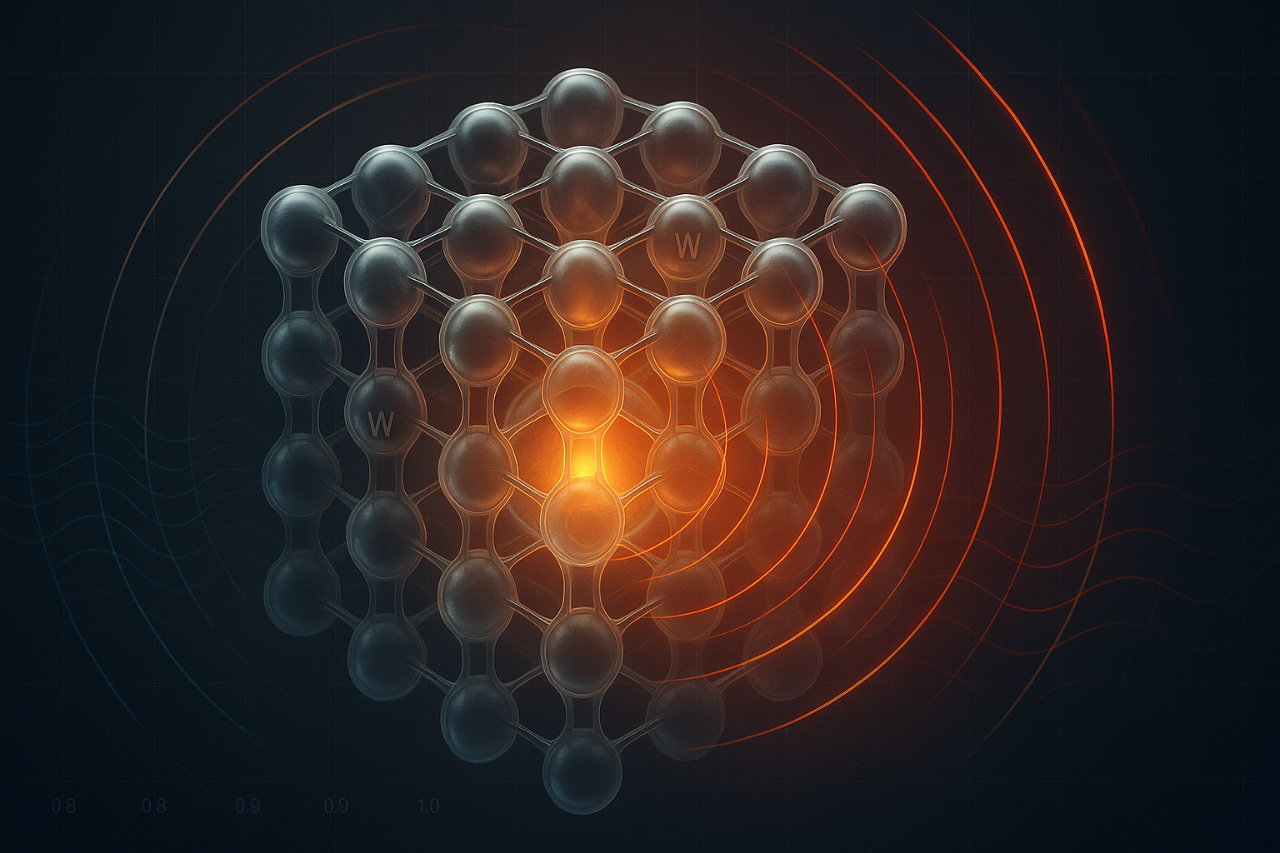
In the study by Arpin et al. (2013), 3D self-assembled tungsten photonic crystals retain their structure up to ~1,400 °C, while exhibiting tailored IR reflectance/emissivity spectra that show enhanced useful thermal emission and suppressed undesired IR emission.
The role of HYPERION: researchers used reflectance IR to infer spectral emissivity via Kirchhoff’s law and applied reflectance analysis to directly link to emissivity control of tungsten photonic crystals.
Nanophotonics / Bioanalytics
Amenabar et al. (2013) demonstrated infrared nanospectroscopy (nano-FT-IR) mapping of individual protein complexes (e.g. ferritin, insulin fibrils) with ~30 nm lateral resolution, interpreting local broadband spectra in the amide I/II region to deduce secondary structure at the nanoscale.
The role of HYPERION: FT-IR spectra were acquired as reference spectra and to compare peak positions and band shapes with the nano-FT-IR data, thereby supporting assignments of local secondary-structure resonances.
Plastmonics / Supercrystals
Mueller et al. synthesized three-dimensional, self-assembled gold nanoparticle “supercrystals” and demonstrated that plasmon polaritons in these crystals induce strong near-field enhancement across visible to mid-IR bands, enabling both SERS and SEIRAS of ligand molecules.
The role of HYPERION: IR reflectance and transmittance spectra on plasmonic supercrystal films where collected demonstrating coupling between plasmonic modes and molecular vibrations and quantifying SEIRAS enhancement.
On-Chip-Screening / Biomedical

In Benz et al. (2020) a dendrimer-based, patterned indium-tin-oxide (ITO) slide was used to generate >50,000 nanodroplet vessels per plate, enabling on‐chip chemical synthesis and ultra-sensitive MALDI-TOF MS, UV–Vis reaction monitoring (RM) and on-chip IR spectroscopy for RM and characterization in high-throughput drug discovery.
The role of HYPERION: IR spectra from the droplet array spots provided molecular vibrational absorption signatures to complement MS and UV-Vis data, thus supporting high-throughput characterization of the synthesized library.
Plasmonics / Metamaterials
Paggi et al. (2023) demonstrate an over-coupled metasurface resonator that enhances molecular vibrational absorption across the mid-IR range (5–10 μm) by coupling weak plasmonic modes to molecular vibrations, achieving reflectivity variations up to 1% per nanometer of molecular layer thickness.
The role of HYPERION: IR spectra in the mid-IR allowed to assess the influence of the metasurface resonator on vibrational modes, shed light onto the broadband SEIRA mechanism and supported the design of detection platforms.
Polariscopy / Nanomaterials

Honda et al. (2019) demonstrated infrared (IR) polariscopy using hyperspectral imaging with a focal plane array (FPA) detector in the IR spectral region under illumination by thermal and synchrotron light sources. They show that the time required for polariscopy at IR wavelengths can be substantially reduced by the FPA-FTIR imaging.
The role of HYPERION: Both the offline FPA-FT-IR experiments and the synchrotron based hyperspectral imaging were performed on the Hyperion enabling the analysis of the targeted sub-wavelength features via polariscopy.
Energy Materials / Metamaterials

Dyachenko et al. report the design and fabrication of a refractory multilayer metamaterial composed of tungsten and hafnium dioxide that exhibits an optical topological transition between epsilon-near-zero and hyperbolic dispersion. This transition allows control over the material’s thermal emission properties at high temperatures, enabling selective tuning of emissivity.
The role of HYPERION: Reflectance spectra were collected in the NIR region to determine the spectral position and strength of optical resonances in the multilayer structure.
Optics / IR Imaging
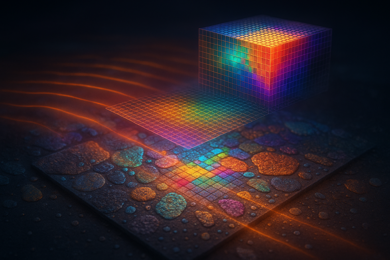
Stavitski et al. (2013) achieved diffraction-limited spatial resolution by coupling synchrotron infrared (IR) beamlines to focal plane array detectors (FPA), enabling full-field imaging with high chemical sensitivity and rapid acquisition times. Using this approach, they successfully map chemical composition in heterogeneous samples with spatial resolution down to approximately 5 µm.
The role of HYPERION: Equipped with an FPA detector, the HYPERION served as the imaging platform coupled to the synchrotron which was used for the chemical and compositional analysis.
Catalysis / Energy Materials

Cao et al. report using operando synchrotron radiation FT-IR (SR-FT-IR) under working potentials to detect a reversible vibrational absorption band at ~764 cm⁻¹ of an atomically dispersed Ru₁–N₄ catalyst, which they attribute to the adsorption/desorption of an oxygen intermediate (O*), thereby revealing the dynamic involvement of O species at the Ru site during catalysis.
The role of HYPERION: An operando SR-FT-IR setup was used to spatially and spectroscopically interrogate the catalyst under bias, capturing the vibrational signature of O* on the Ru site and supporting direct linkage between intermediate adsorption and catalytic function.
Catalysis / Energy Materials

In Su et al., the authors design CuO clusters supported on N-doped carbon and show that under applied potential the clusters dynamically reconstruct into Cu₂–CuN₃ species with charge asymmetry, which correlate with high Faradaic efficiency toward C₂⁺ products (notably ethanol). They combine operando XAS, quasi-in situ XPS, and operando FT-IR (with DFT support) to trace structural, electronic, and adsorbate changes in real time.
The role of HYPERION: As part of their operando FTIR setup, it was used to focus the synchrotron-based IR beam into the reaction cell and collect reflectance spectra under applied potentials. These measurements enable tracking of molecular vibrational features and charge-asymmetry signatures linked to the Cu₂–CuN₃ clusters and thus substantiate the structure–activity correlations under working conditions.
IR Sampling Accessories and Objective Lenses
Validation Plate

MicroVice Sample Holder

ATR Macro Imaging Accessory

Filter Holder

Heating and Cooling Stage

3.5x Imaging Objective Lens
Standard Transmission Objective Lenses

ATR Objective

Grazing Angle Objective

Diamond Compression Cell

OPUS Release 9.0 Highlights for HYPERION II | Q1 2025
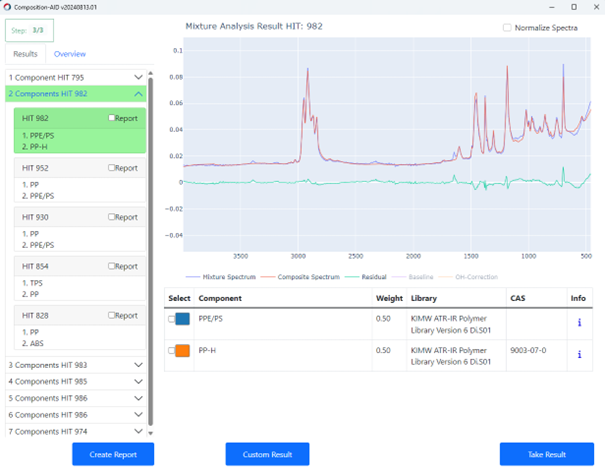
New Feature: The Autonomous Composition Identifier (A.I.D.) simplifies search and identification.
The Autonomous Composition Identifier (A.I.D.) is an AI-driven software tool for identifying the chemical composition of samples based on IR or Raman spectral data. It incorporates the previous O/SEARCH features and adds a multi-step algorithm to find the best match from reference spectral databases, whether dealing with pure substances or complex mixtures. A.I.D. intelligently evaluates and recommends optimal matches with a balance of reliability and sensitivity—automatically generating diagnostics and visual reports, including residual spectra and hit quality graphs. It delivers high-performance results in seconds and supports in-depth, interactive analysis of alternative identifications.
New Feature: Co-local Measurement – Unified Analysis Across IR and Raman
The Co-local Measurement function, part of the OPUS/OBJECT package, enables seamless analysis of the same region of interest on microscopic samples using both IR and Raman microscopy. It allows the transfer of overview images and measurement areas between LUMOS II, HYPERION II, and SENTERRA II instruments. This ensures that IR and Raman data are collected from precisely the same spot, enabling complementary chemical insights. Co-local Measurement bridges the gap between two techniques, improving data correlation and boosting confidence in results for heterogeneous or complex samples.

Updated Feature: OPUS/OBJECT now combines "Find Particle" and "Cluster ID"
The Particle Identification function (part of the OPUS/OBJECT package) combines the power of “Find Particles” and “Cluster ID” into one streamlined workflow. It detects particles within IR and Raman chemical images, determines their dimensions, and identifies their chemical makeup—all in one go. This integrated approach simplifies analysis and accelerates decision-making for complex samples.

OPUS Release 8.7 | HYPERION II | Q3 2021
New Feature: High Performance Chemical Image Generation by New Adaptive K-means Clustering Function
This new function is the logical next development step for our well known Cluster analysis function.The Adaptive K-means Clustering Function is based on a new algorithm, which enables a non-supervised and autonomous determination of spectral variance within your imaging or mapping results.
- Forecasting or time consuming searching of the included amount of chemical classes is no longer been necessary as the algorithm can predict all included chemical classes by itself.
- This major function is important for all kind of chemical imaging and distribution analysis of unknown samples or small structures within larger datasets.
- Make analysis and evaluation is as easy as possible and safes your valuable time and nerves.
New Feature: “Cluster ID” Function for Identification of Classes in 3D Spectral Data
Our new Cluster ID function enables the identification of clusters within imaging and mapping data using the OPUS functions: spectrum search in libraries, quick compare, or identity test.
- Easy determination of the chemical identity of classified sample components for particles, layers in laminates, components of pharmaceutical tablets and other inhomogeneous materials.
- Reliable and comprehensive statistics reports about quantity, size and of course identity of all analyzed structures is provided and leads particle and technical cleanliness analysis to a new, autonomous level.
Updated Feature: "Find Particles" function now contains a novel particle detection method
The proven "Find Particle" software can now be applied to both: the visual and the IR image. With this updated feature, you are able to do particle detection based on chemical images that were measured by the LUMOS II.
- While particle recognition for low contrast structures and off-white/transparent particles/fibers on off-white filter membranes can be tedious, a postrun particle determination based on the chemical IR image allows you to determine quantity and size of particles from your imaging or mapping results.


Literature Room
Learn more about our FT-IR microscopes and solutions by downloading related literature.


